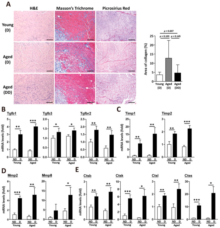
Figure 4.
Upregulation of TGF-β signaling activated ECM remodeling after muscle injury. (A) Histological results of young and aged skeletal muscle after muscle injury, including H&E staining, Masson’s trichrome staining, and picrosirius red staining. Muscle in the aged double dose (dd) group was damaged twice at day 0 and day 7. Quantitative data of collagen area (blue part) from Masson’s Trichrome staining results in young and aged skeletal muscle. At least 5 captured images of muscle sections from different fields in each mouse sample were examined, and there were 3 mice from each group. Scale bar, 100 µm. (B) Quantitative data of RT-qPCR mRNA expression of TGF-β, (C) Timp genes, (D) Mmp genes, and (E) cathepsin genes in young and aged skeletal muscle. There were 4 mice in each group. ND, nondamaged muscle as a control. D, Fourteen days after BaCl2 muscle injury. The results are presented as the mean ± SD. * p < 0.05; ** p < 0.005; *** p < 0.001.

