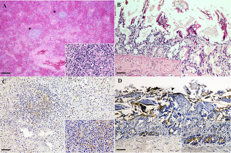
Fig 1. CPPV-1 infection in fishing cats.
Demonstrative H&E (A, C) and CPPV-1 IHC (B, D) pictures from fishing cats. (A) Fishing cat no. 1. Diffuse congested spleen with sparse numbers of lymphoid follicles. Center of one of the remaining lymphoid follicles contained eosinophilic fibrillar material (fibrin) intermixed with scattered karyorrhectic debris of lymphocytes (lymphocytolysis) (inset). Few numbers of these lymphocytes contained 5–7 μm basophilic intranuclear inclusion bodies that marginated the nuclear chromatin (arrow). (B) Fishing cat no. 3. Shortening of villi with occasional dilated crypts that contained eosinophilic proteinaceous substances and lined by markedly attenuated or necrotic crypt epithelial cells (inset). Many crypt epithelial cells were pyknotic and karyorrhectic and rare cells contained similar basophilic intranuclear inclusion bodies (arrows). (C) Fishing cat no. 1. The CPPV-1 immunoreactivity was frequently observed in the cytoplasm of mononuclear cells in the area of splenic lymphoid follicle. (D) Fishing cat no. 3. CPPV-1 IHC signals were diffusely detected and the immunoreactivity signals were markedly localized in the cytoplasm of cryptal epithelial cells (inset). Bars indicate 25 μm for (A) and 120 μm for (B–D).

