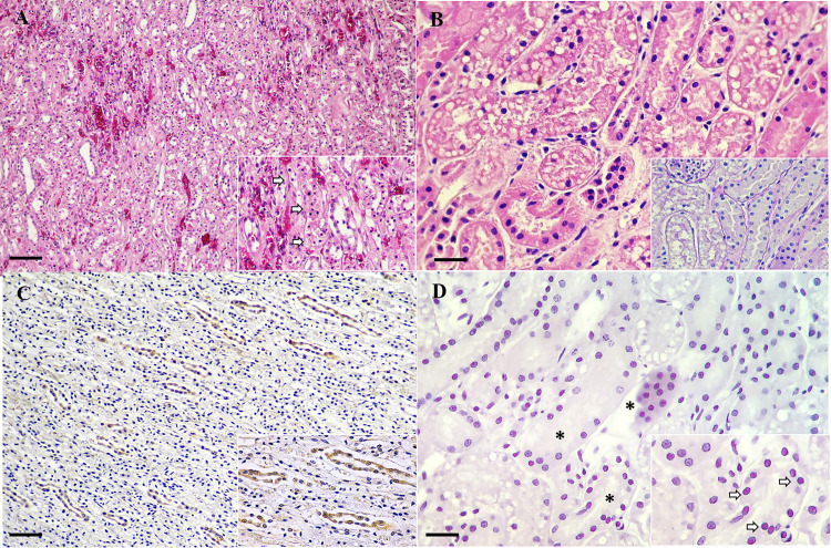
Fig 2. CPPV-1 infection in fishing cats.
Demonstrative H&E (A), PAS staining (B), CPPV-1 IHC (C) and in situ hybridization (ISH) (D) photomicrographs of kidney from fishing cats. (A, B) Fishing cat no.3. (A) Multifocal renal hemorrhage with renal tubular vacuolation. Few tubular epithelial cells were hypereosinophilic with pyknotic nuclei (inset, arrows). (B) Renal tubular epithelial cells exhibited cytoplasmic vacuole and the nuclei of rare epithelial cells are vesiculate with marginating nuclear chromatin. PAS staining highlighted intact basement membrane (inset). (C) Fishing cat no. 2. CPPV-1 IHC-immunoreactivity (dark-brown color) was diffusely observed in the renal tubules and frequently detected in the cytoplasm of renal tubular epithelium (inset). (D) Fishing cat no.1. CPPV-1 ISH-immunoreactivity (reddish pink color) was abundantly observed in renal tubules (asterisks), and it localized in the nuclei of renal tubular epitheliums (inset, arrows). Bars indicate 50 μm for (A, C) and 120 μm for (B, D).

