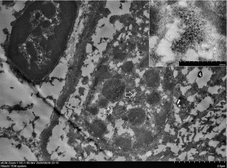
Fig 3. CPPV-1 viral particles in the nucleus of renal tubular epithelial cells.
Transmission electron microscopy (TEM) using the pop-off technique. Ultrastructural demonstration of clustering of electron-dense particles (arrows). The icosahedral particle size was 19–22 nm diameter located in the nucleus of renal tubular epithelial cells (inset). Scale bars as shown in the figure.

