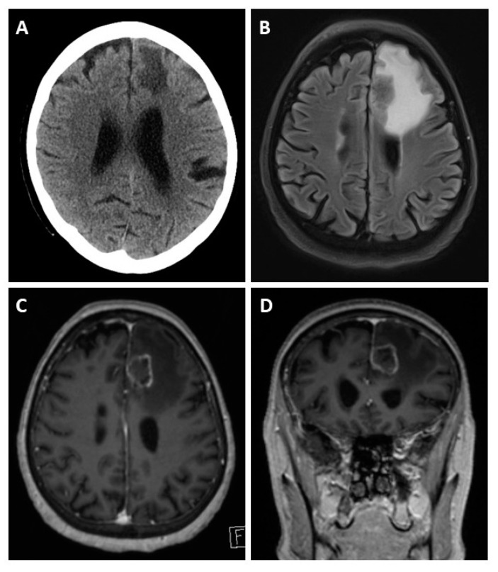
Figure 3.
Cerebral CT and MRI scans of a patient with BM, a 62-year-old patient presenting with left frontal edema in computed tomography (A). The MRI scan reveals the actual extent of the edema in the fluid-attenuated inversion recovery (FLAIR) sequence (B). T1-weighted gadolinium post-contrast images in the axial (C) and coronal (D) orientation reveal causative left-frontal CRC metastasis.

