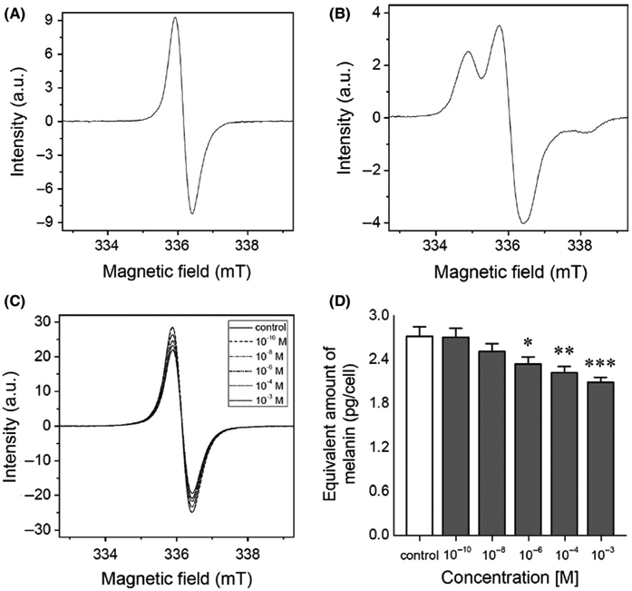
FIGURE 2.

Melatonin decreases melanin content in MNT-1 melanoma cells. Based on assessment carried out by electron paramagnetic resonance (EPR) spectroscopy, here the spectra of (A) synthetic DOPA-melanin used as standard for melanin determination; (B) synthetic cysteine-L-DOPA melanin indicating the characteristic of EPR signal of pheomelanin, and cells after 72 h incubation with melatonin in dose-dependent manner are depicted (C). Bar graph displays mean values + SD (n = 3) of pg of melanin per cell in melatonin-treated cells (D). Statistically significant differences versus control were indicated as *P < .05, **P < .01, ***P < .001
