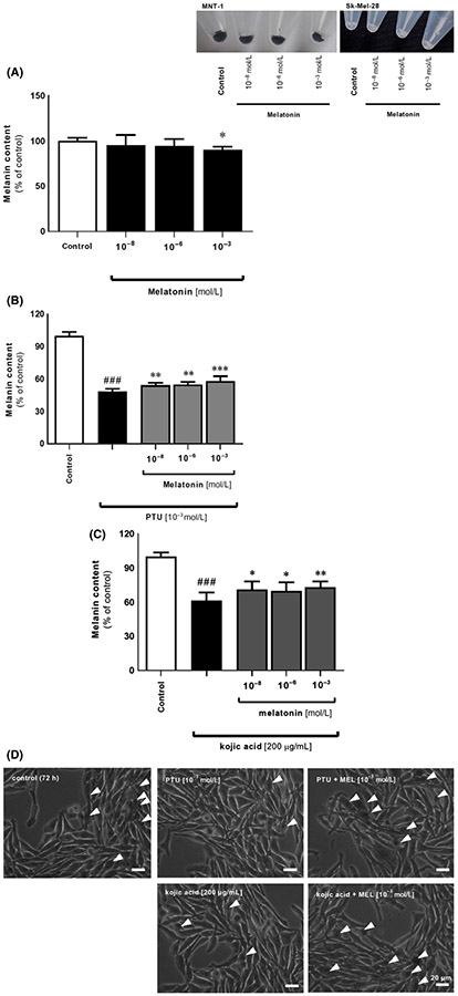
FIGURE 3.
Melatonin counteracts the inhibitor-mediated alterations in cell pigmentation in MNT-1 cells. Images enclosed as insert present distinct differences in pigmentation ratio between MNT-1 and Sk-Mel-28, representatives for highly pigmented and amelanotic cells, respectively. (A) Evaluation of melanin content has been carried out using melanotic MNT-1 cells cultured in MEM supplemented medium for 72 h with melatonin (10−8, 10−6, 10−3 mol/L) and melanogenic inhibitors, that is, 10−3 mol/L PTU (B) or 200 μmol/L kojic acid (C) as described in Materials and Methods. Data were presented as the mean + SD (n = 5), the values are expressed as percentage of the control sample. Statistically significant differences were indicated as *P < .05, **P < .01, ***P < .001 while comparison of PTU- or kojic acid-treated cells versus control sample was indicated as ###P < .001. (D) Visualization of MNT-1 cells in culture and effect of melatonin versus PTU or kojic acid. Arrowheads point out pigmented cells

