. 2021 Mar 1;10:e65339. doi: 10.7554/eLife.65339

Figure 4. Structural transitions around the aromatic rich loop.

(A) Molecular secondary structure cartoons for the region surrounding the aromatic rich loop (ARL) of BLM-HDΔWHD (top), PDB entry 4CGZ; BLM-HD in complex with DNA (middle) and liganded complex; BLM-HDΔWHD in complex with ADP, ssDNA-15mer and 2 (bottom). The side chains for key amino acid residues are shown in stick representation, with carbon atoms coloured according to their respective domains (see associated key). Bound ADP and 2 are also shown in stick representation, with carbon atoms coloured grey and yellow, respectively. (B) Expanded and rotated view highlighting the interactions made between the ARL and ssDNA-15mer oligonucleotide (cartoon coloured cyan) in the liganded complex, also showing the relative position of compound 2. Potential hydrogens bonds are represented by black dotted lines.

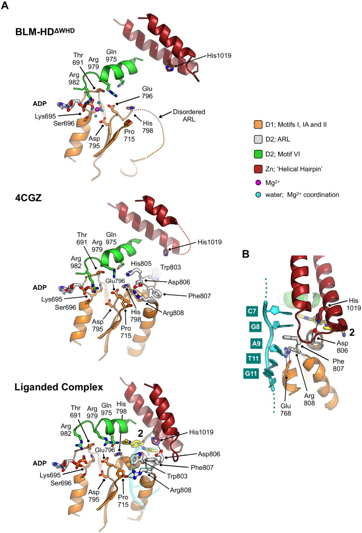
Figure 4.

Figure 4—figure supplement 1. Molecular secondary structure cartoons showing selected amino acid side chains of the aromatic-rich loop (ARL) region in PDB entries 4CDG and 4CGZ (BLM-HD) and 4TMU (Cronobacter sakazakii RecQ) to that reported here for liganded-BLM-HDΔWHD.

Figure 4—figure supplement 1.

Amino acid numbering is provided for the start and end of the region compared, with the identity of key residues also provided.
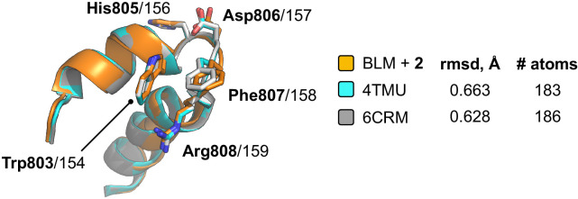
Figure 4—figure supplement 2. Superposition of the ARL in liganded-BLM-HDΔWHD (coloured orange) with those found in PDB entries 4TMU (cyan) and 6CRM (Voter et al., 2018) (grey); which both represent structures of the catalytic core of C. sakazakii RecQ in complex with different DNA substrates.

Figure 4—figure supplement 2.

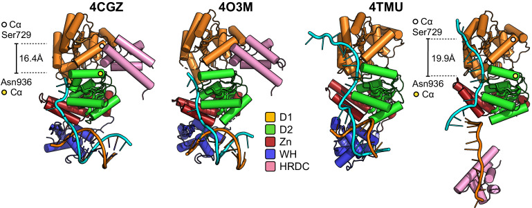
Figure 4—figure supplement 3. Molecular cartoon representations of the helicase catalytic cores reported in PDB entries 4CGZ, 4O3M, and 4TMU, highlighting their respective interactions with bound DNA substrates and comparing this to liganded BLM-HDΔWHD.

Figure 4—figure supplement 3.

Additional details for the colour scheme can be found in the associated key. The distance between the C-alpha positions of Ser729 and Asn936 is also shown, highlighting a widening of the pocket in our liganded complex (and 4TMU) that would normally serve to accept the HRDC in its ‘parked’ position (4CGZ/4O3M).