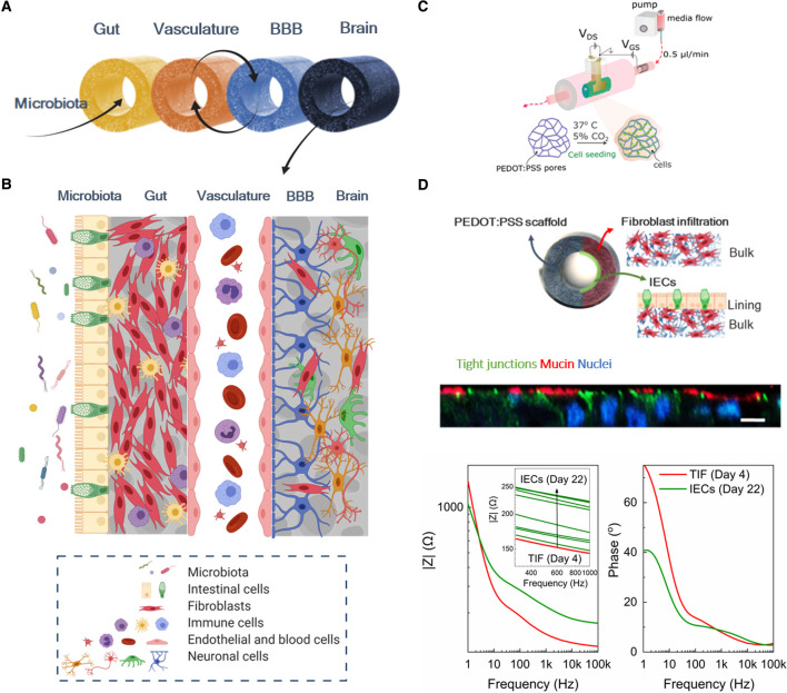
Figure 3. Design and tools of the IMBIBE platform.
(A) Modules of the microbiota-gut-brain axis segments are built to operate independently but can also be interconnected to mimic the in vivo situation, by means of fluidic coupling and interconnection of each tissue equivalent via their bulk compartments; (B) Illustration of the biological components of the complete IMBIBE platform; Created with BioRender.com (C) Schematic of the structure and setup of the ‘Tubistor’, the novel 3D bioelectronic device for building each module of the IMBIBE platform. Reproduced from [109] under the Creative Commons License. (D) The gut module of the IMBIBE platform, hosted in the new generation of the device in (C) — the ‘L-Tubistor’ — modified to better capture the native tissue architecture. A schematic illustration of the intestinal model showing the organisation of different cell components in the hollow tubular electroactive scaffolds of the L-Tubistor (top). Snapshot of z-stacked confocal images illustrating the brush border of the polarised intestinal epithelial layer on the scaffold lumen lining (middle; scale bar 20 µm). Representative graph of electrical monitoring of the intestinal model showing the response of the electroactive scaffolds to tissue formation from day 4 of fibroblast culture to day 22 of intestinal cell culture (overall day 26; bottom). Adapted from [110] under the Creative Commons License.

