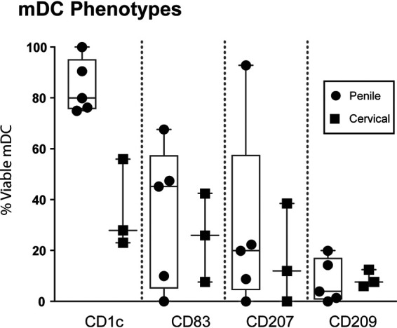
FIG 2.

Characterization of the migratory cell phenotype in penile and cervical tissue. The percentages of CD1c-, CD83-, CD207-, and CD209-positive cells of all viable mDCs isolated from penile (n = 5) and cervical (n = 3) tissue were assessed by a multicolor flow panel on an LSRIIFortessa flow cytometer. Box-and-whisker plots show median, minimum/maximum, and 25th/75th percentiles of the percentage of viable cells stratified by CD1c, CD83, CD207, and CD209 expression. Each symbol represents an individual penile or cervical tissue donor.
