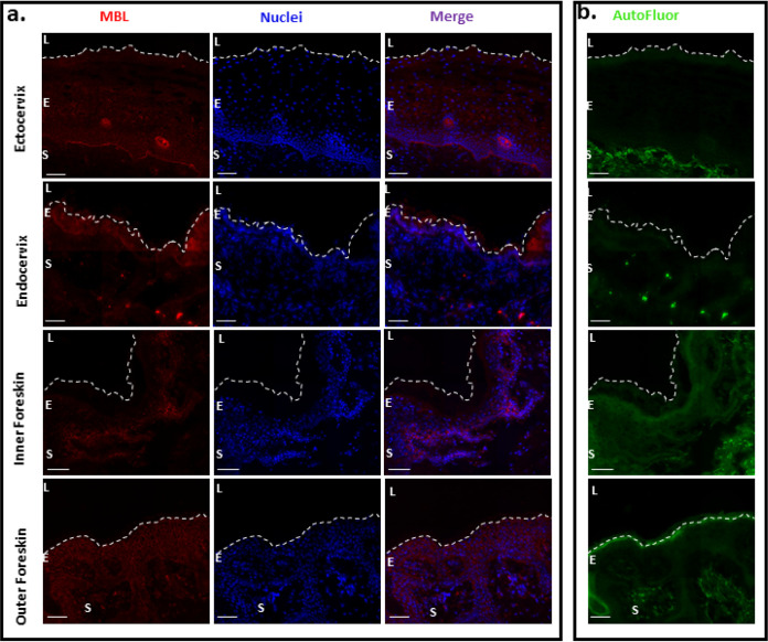
FIG 7.
Presence of MBL in different mucosal tissue types. (a) Fluorescent deconvolution microscopy images of ectocervix, endocervix, inner foreskin, and outer foreskin stained for MBL (red) and DAPI (blue). (b) Autofluorescence (green) of the corresponding tissues. Magnification, ×40. Bar, 40 μm. Dashed lines indicate the luminal surface. L, lumen; E, epithelium; S, stroma.

