FIG 3.
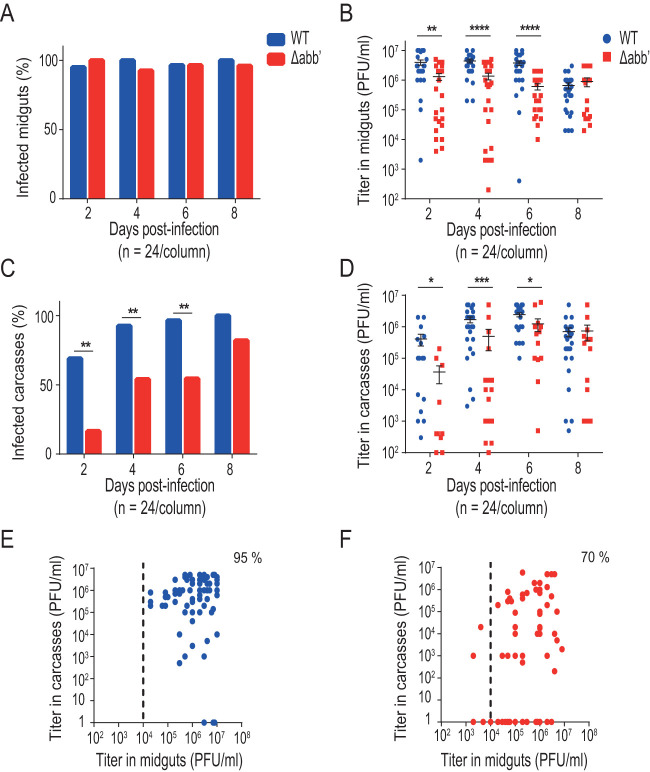
Increasing the infectious dose decreases the midgut escape barrier effect. Mosquitoes were blood fed with 5 × 10⁶ PFU/ml wild-type (WT) or Δabb′ mutant CHIKV and dissected from days 2 to 8 to separate midguts and carcasses. Infection rates and viral titers were measured in each sample. (A) Bar graph showing midgut infection rates. (B) Dot plot showing mean viral titers and SD of WT and Δabb′ viruses in midguts of infected mosquitoes. (C) Bar graph showing carcass infection rates. (D) Dot plot showing mean viral titers and SD of WT and Δabb′ viruses in carcasses of infected mosquitoes. (E and F) Scatterplot of viral titers in the midgut versus carcass for wild type (E) and mutant (F) viruses. Statistics on infection rates were performed by Fisher’s exact test on cumulative data (n = 24) from two independent experiments. Statistics on viral titers were performed by a Mann-Whitney U test.

