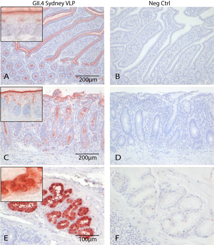
FIG 1.
GII.4 VLPs attached to epithelium of villi and crypts in human small intestinal tissues (red). No difference was seen between FITC-labeled (A) and unlabeled (C) VLPs that were detected with an anti-FITC and an anti-GII.4 antibody, respectively. (E) In some tissues, VLPs additionally attached to the Brunner glands. (B, D, and F) No staining was seen in the negative controls (Neg Ctrl). Magnifications, 20× (A, B, C, and D) and 20× (E and F). Picture insets of attachment signal are 100×.

