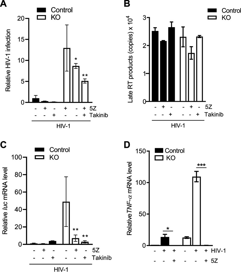
FIG 5.
TAK1 inhibition attenuates HIV-1 infection in THP-1 cells lacking SAMHD1 expression. (A) THP-1 cells were treated with 5Z (1 μM) for 30 min, Takinib (10 μM) for 2 h, or DMSO. Inhibitor-containing medium was removed prior to 2 h infection with single-cycle HIV-1-Luc/VSV-G (MOI, 1). Takinib was added and maintained in culture for 24 h postinfection (hpi). Cells were harvested at 24 h for luciferase assay. All luciferase values were normalized to 10 μg protein. Relative HIV-1 infection was calculated by setting mock-treated cells as 1. Data represent 4 replicates, and error bars show SEM. (B) At 6 hpi, late reverse transcription (RT) products were quantified by qPCR assays using samples from the experiment in panel A. Serial dilutions 108 to 101 of a proviral pNL4-3 plasmid were used to calculate copy numbers of late RT products. Each biological sample was run in duplicate, and unspliced GAPDH was used for normalization. (C) Measurement of mRNA levels was performed from samples in the same experiment described in panel A. Cells were harvested at 18 hpi for luc mRNA quantification by RT-qPCR. 18S rRNA was used as a normalization control. The graph depicts data derived from triplicate samples with error bars representing SEM. (A and C) Statistical analysis was performed by two-way ANOVA with Tukey’s multiple comparisons posttest. (D) Cells were treated with 5Z (1 μM) for 30 min or DMSO. Cells were infected for 2 h with HIV-1-Luc/VSV-G (MOI, 2) in the presence of the inhibitor. TNF-α mRNA levels at 2 hpi were quantified by RT-qPCR and normalized to spliced GAPDH. Error bars represent SD of triplicate samples. Statistical significance was calculated by unpaired t test. *, P ≤ 0.05; **, P ≤ 0.01; ***, P ≤ 0.001.

