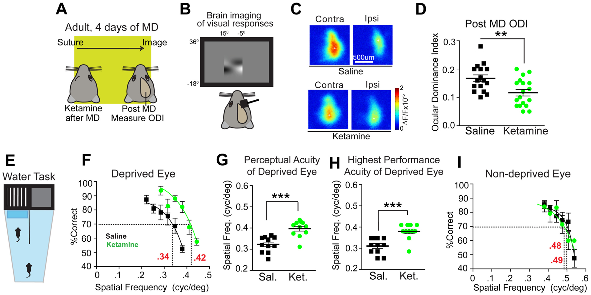
Figure 1. Ketamine reactivates adult ocular dominance plasticity and restores visual acuity in amblyopic animals.

(A-D) Testing the effect of ketamine on ocular dominance plasticity (ODP) during adulthood using intrinsic signal optical imaging. (A) Illustration of the experimental paradigm in adult (P90–120) mice, and the timeline of the protocol. (A, left) After monocular deprivation (MD), saline or ketamine treatments are given, and 4 days later ODP is measured after eyelid suture removal. (B) Intrinsic signal responses of visual cortex to contralateral versus ipsilateral eye stimulation are recorded and the ocular dominance index (ODI) is assessed. The ODI is computed as (C-I)/(C+I) where C and I are the averaged map amplitudes calculated for contralateral and ipsilateral visual stimulation. (C) Representative cortical response maps from a control saline treated animal (top) and a ketamine treated animal (bottom) 4 days after treatment. In adult mice, 4 days of MD does not change ODIs. (D) However, ODI is reduced in ketamine treated adult animals (n=18, green) as compared to saline treated animals (n=16, black) (Mann-Whitney U test, p = 0.0078; mean ± SEM). The data were pooled from cohorts of mice receiving either 10 or 50 mg/kg ketamine treatment, as they shared similar trends. (E-I) Testing the effect of ketamine on restoring functional visual perception in adult amblyopic mice. (E) The visual water maze task (VWMT). Mice had one eye sutured shut from P18-P32. At P32 the eye was re-opened and mice were treated with ketamine (10mg/kg; s.c.) every other day for three total treatments. At P90 mice began training, and were tested at P100. Testing continued until ~P140, with mice doing ~10 trials/day. (F) Performance on the VWMT for all mice treated with either saline (black, n=11) or ketamine (10mg/kg; s.c., green, n=12) using the deprived eye. The performance curve is fit to a sigmoid using the last 6 data points [34]. The x-axis represents the spatial frequency of the visual stimulus gratings. The threshold of perceptual acuity is determined by the spatial frequency (x-axis) corresponding to the 70% value (y-axis, dashed line). For controls the acuity threshold is 0.34 cpd. For ketamine the acuity threshold is 0.42 cpd. (G) Perceptual acuity of deprived eyes across mice using acuity thresholds determined from performance curves of individual eyes. For deprived eyes ketamine (green, n=12) shows significantly improved perceptual acuity compared to saline (black, n=11) (Mann-Whitney U test, p = 0.0008; mean ± SEM). (H) Highest performance acuity at or above the 70% level for the deprived eye on the VWMT for mice treated with saline (black, n=11) or ketamine (green, n=12). Performance acuity is significantly increased with ketamine (Mann-Whitney U test, p = 0.0009; mean ± SEM). (I) Average performance on the VWMT for mice treated with saline (black, n=11) or ketamine (green, n=12) using the non-deprived eye. For saline the acuity threshold is 0.49 cpd. For ketamine the acuity threshold is 0.48 cpd. Average perceptual acuity between groups determined from performance curves of individual non-deprived eyes yielded no significant change (data not shown; Mann-Whitney U test, p = 0.1927; mean ± SEM). Also see related information in Figure S1 and Table S1.
