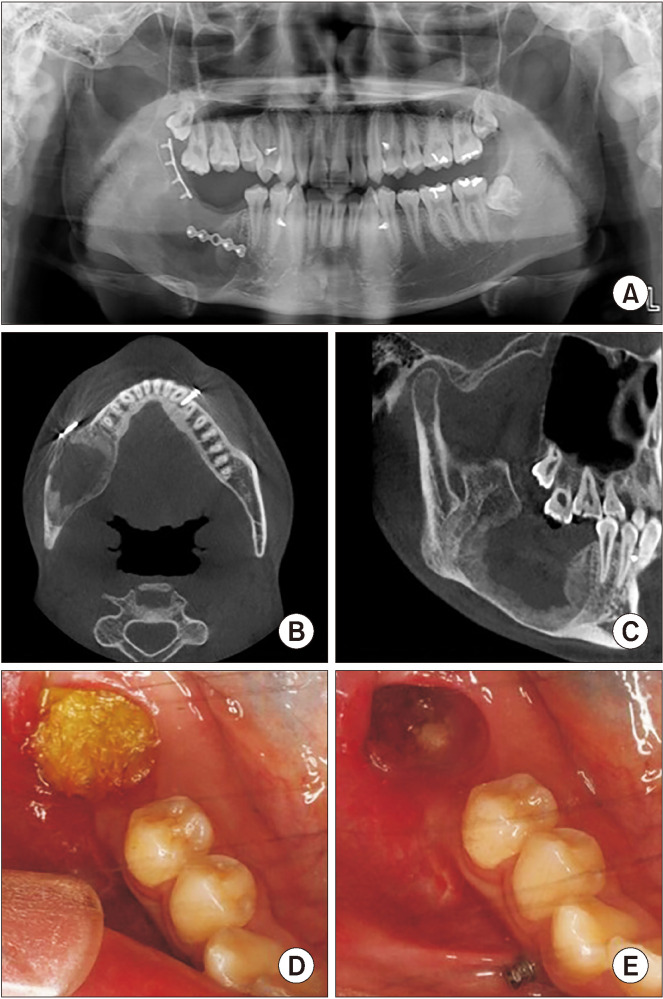
Fig. 9.
Radiograph after 22 weeks of marsupialization. A. Panoramic view on #45 distal, posterior, and inferior borders of the lesion: radiopacity was increased. B. Coronal view on computed tomography (CT): new bone formation was observed along the mass removal area. C. Sagittal view on CT: new bone formation was observed along the mass removal area. D. Intraoral photograph at 22 weeks of cyst enucleation: furacin gauze was changed every week. E. Soft tissue healing was observed after 22 weeks of cyst enucleation.

