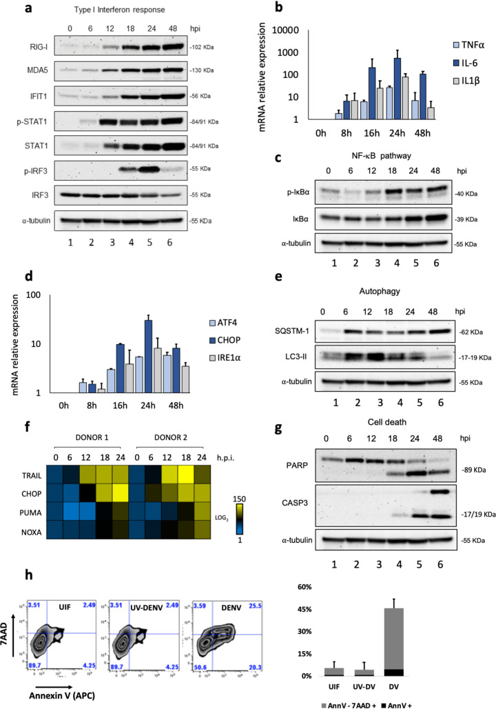
FIG 1.
DENV-associated host innate immune and stress responses in Mo-DC. Human Mo-DC were infected with DENV at an MOI of 1, and samples of cells were collected at 0, 6, 12, 18, 24, and 48 hpi. (a) Protein analysis by Western blotting of primary Mo-DC treated with DENV-2 (MOI, 1). Detection of endogenous human RIG-I, MDA5, IFIT1, STAT1, and IRF3 proteins was used to address the activation of the type I interferon pathway. An anti-α-tubulin antibody was used to measure total loaded protein in the Western blot. Results for all Western blots are representative of two independent experiments. (b) TNF-α, IL-6, and IL-1β gene expression levels were evaluated by qPCR analysis; results are representative of three independent experiments and are expressed as means ± SD from three biological replicates. (c) Antibodies against phosphorylated IκBα or total IκBα (Cell Signaling) were used to determine the levels of IκBα and phospho-IκBα, as a measure of NF-κB activation. (d) ATF4, CHOP, and IRE1α gene expression levels were evaluated by qPCR analysis. (e) Antibodies against SQSTM-1 and LC3-II were used as markers for the induction of autophagy. (f) High-throughput qPCR Biomark analysis of the expression of selected apoptotic genes. Gene expression levels were calculated using the ΔΔCT method. The scale represents the log2 values, where yellow shows upregulation, and blue shows downregulation, in gene expression. Data are representative of one experiment performed on two individual donors (donor 1 and donor 2). (g) PARP and caspase-3 antibodies were used as measures of cell death. (h) Apoptosis was quantified by combined staining with annexin V and 7-AAD, and fluorescence was analyzed at 48 hpi using flow cytometry. Percentages are shown for early apoptotic cells (annexin V positive) and late apoptotic/necrotic cells (positive for both annexin V and 7-AAD). UIF, uninfected. Data are representative of the results of three independent experiments.

