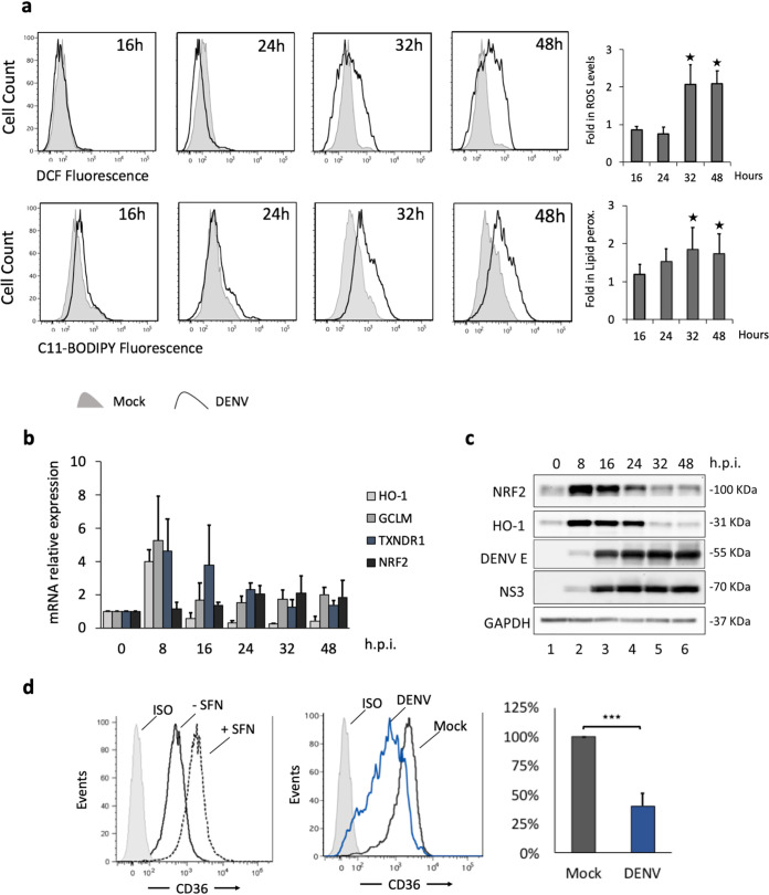
FIG 2.
DENV increases oxidative stress in primary human DC. Human Mo-DC were either infected with DENV-2 NGC at an MOI of 1 or mock infected, and cells were collected at 16, 24, 32, and 48 hpi. (a) (Top) The accumulation of ROS was measured by flow cytometry using CM-H2DCFDA (1 μM) at the indicated times. (Bottom) Lipid peroxidation was measured by using C11-Bodipy (1 μM). Data represent means from at least three different donors. P values and fold increases were determined based on comparison with uninfected cells. (b) HO-1, GCLM, TXNDR1, and NRF2 gene expression levels were evaluated by qPCR analysis, normalized to GAPDH expression, and described relative to control levels (0 hpi). Results are representative of three different donors and are expressed as means ± SD. (c) Protein analysis by Western blotting of primary Mo-DC treated with DENV-2 (MOI, 1). Endogenous human NRF2 and HO-1 proteins were detected after 0, 8, 16, 24, 32, and 48 hpi. DENV infection was detected using a polyclonal rabbit antibody raised against the DENV-2 E protein or the DENV-2 NS3 protein. An anti-GAPDH antibody was used to measure total loaded protein in the Western blot. Results are representative of two different donors. (d) (Left) Mo-DC were examined for CD36 expression by flow cytometry after 24 h of treatment with sulforaphane (10 μM). ISO, •••••••. (Center) Mo-DC were infected with DENV at an MOI of 1 or were mock infected, and samples were collected at 40 hpi prior to FACS analysis. (Right) Downregulation of CD36 was measured as a percentage determined based on comparison with levels in mock-infected cells. Data are means of results from three independent donors.

