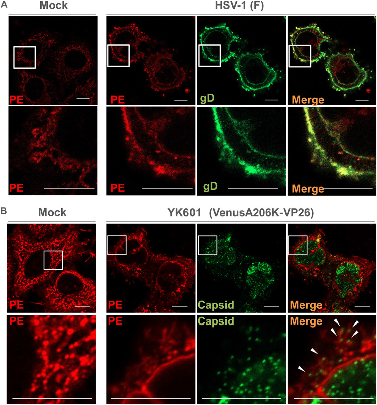
FIG 6.
Subcellular localization of PE in HSV-1-infected cells. (A) HeLa cells were mock infected or infected with wild-type HSV-1(F) at an MOI of 5. At 18 h postinfection, the cells were fixed, permeabilized, and stained with duramycin (red) or anti-gD antibody (green). Each lower image is a magnification of the boxed area in the image above it. Bars, 10 μm. (B) HeLa cells were mock infected or infected with HSV-1 YK601 (VenusA206K-VP26) at an MOI of 5. At 18 h postinfection, the cells were fixed, permeabilized, stained with duramycin, and analyzed with the Airyscan system. Each lower image is a magnification of the boxed area in the image above it. Arrowheads indicate PE-stained structures associated with capsids. Bars, 10 μm.

