FIG 3.
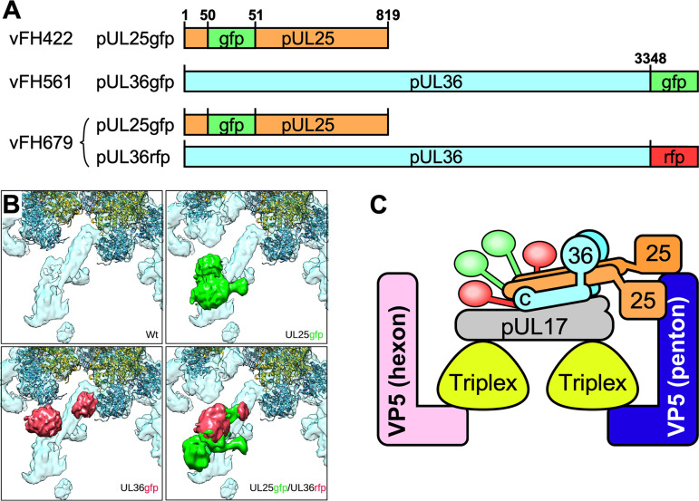
Composition of the CVSC helix bundle probed by cryoEM of fusion protein-labeled capsids. (A) The locations of insertions into the pUL25 and pUL36 sequences are indicated. Note that GFP-labeled pUL36 was used in the single mutant vFH561, but the double mutant vFH679 used RFP-labeled pUL36. (B) Views of the CVSC molecule in capsid reconstructions from virions isolated from Vero cells infected by KOS and each of the three mutants. Additional density in the mutant maps is colored green for the pUL25 label and red for the pUL36 label. Both of the single-mutant maps reveal two extra density regions, indicating that the two copies of the labeled protein, pUL25 or pUL36, are present in each case. The double mutant appears to show the crowding of four labels in close proximity. (C) A model of CVSC organization incorporating two copies each of pUL25 and pUL36 and one copy of pUL17, accommodating the observation that the helix bundle in the CVSC molecule comprises 5 helical rods (20). The fusion labels are colored green (pUL25) and red (pUL36) and indicate the approximate locations of corresponding density in panel B and the positions on the proteins they label.

