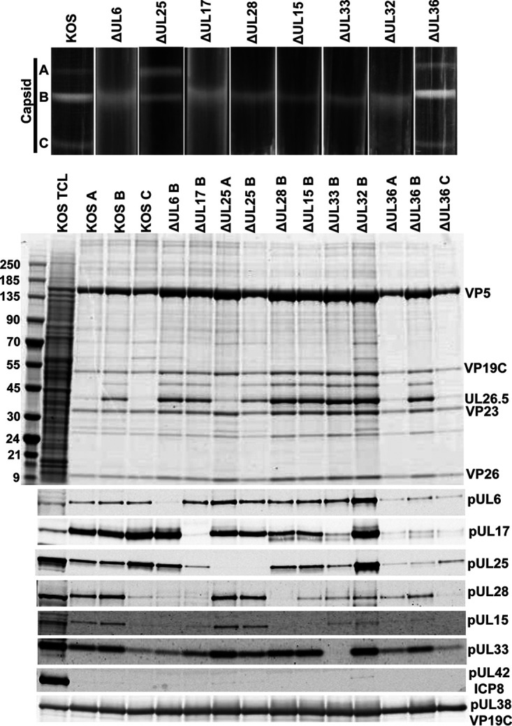
FIG 4.

Analysis of capsid-bound packaging proteins. (Top) Rate-velocity sedimentation of capsids. Cells were infected with KOS or the indicated packaging mutants. Vero cells were infected with 10 PFU per cell for 18 h and nuclear lysates were layered onto 20% to 60% sucrose gradients and centrifuged at 24,000 rpm for 1 h. The positions of A-, B-, and C-capsid bands are indicated. (Center) Coomassie blue-stained gel of capsids (A, B, or C) isolated from KOS and the indicated packaging mutants. Positions of the major capsid protein (VP5), triplex proteins (VP19C and VP23), and scaffold protein (UL26.5) are indicated; KOS TCL, total cell lysate. (Bottom) Western blots of the capsid fractions probed with antibodies for individual packaging proteins. ICP8 (DNA replication protein) and pUL38 (capsid protein) serve as negative and positive controls, respectively. Molecular mass standards (kDa).
