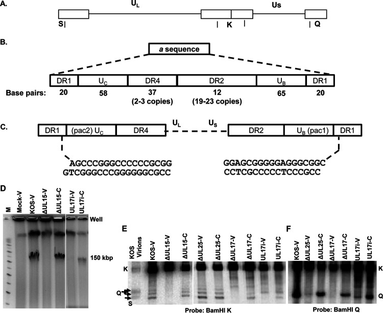
FIG 5.
HSV genome and packaging motifs and processing of virus DNA. (A) Diagram of the 152-kbp HSV-1 genome showing the locations of the BamHI K, Q, and S fragments. (B) Structure of the a sequence elements. (C) The location of the pac1 and pac2 sequences within the UC and UB regions and the sequence of the genome ends, resulting from cleavage within the DR1 element of adjacent a sequences. (D) EtBr-stained PFGE of DNA from mock, KOS, UL15-null, and UL17i virus-infected Vero (V) cells or UL15-null and UL1i-infected complementing (C) cells. (E and F) Well DNA isolated following PFGE (E) or total infected cell DNA isolated from Vero (V) or complementing (C) cells (F) was digested with BamHI followed by Southern blot analysis with 32P-labeled BamHI K (E) or the BamHI Q (F) fragments shown in panel A. The different sizes of the BamHI S fragment are due to the presence of one or more copies of the a sequence present at the UL terminus.

