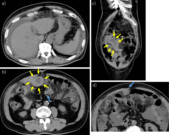
Figure 1.
Abdominal computed tomography. a) The massive ascites. b) The localized small intestinal wall thickening, which is the primary lesion of diffuse large B-cell lymphoma (yellow arrows). There were some intraperitoneal nodules (blue arrow). c) The coronal section at the localized small intestinal wall thickening lesion (yellow arrows). d) There were some intraperitoneal nodules (blue arrow).

