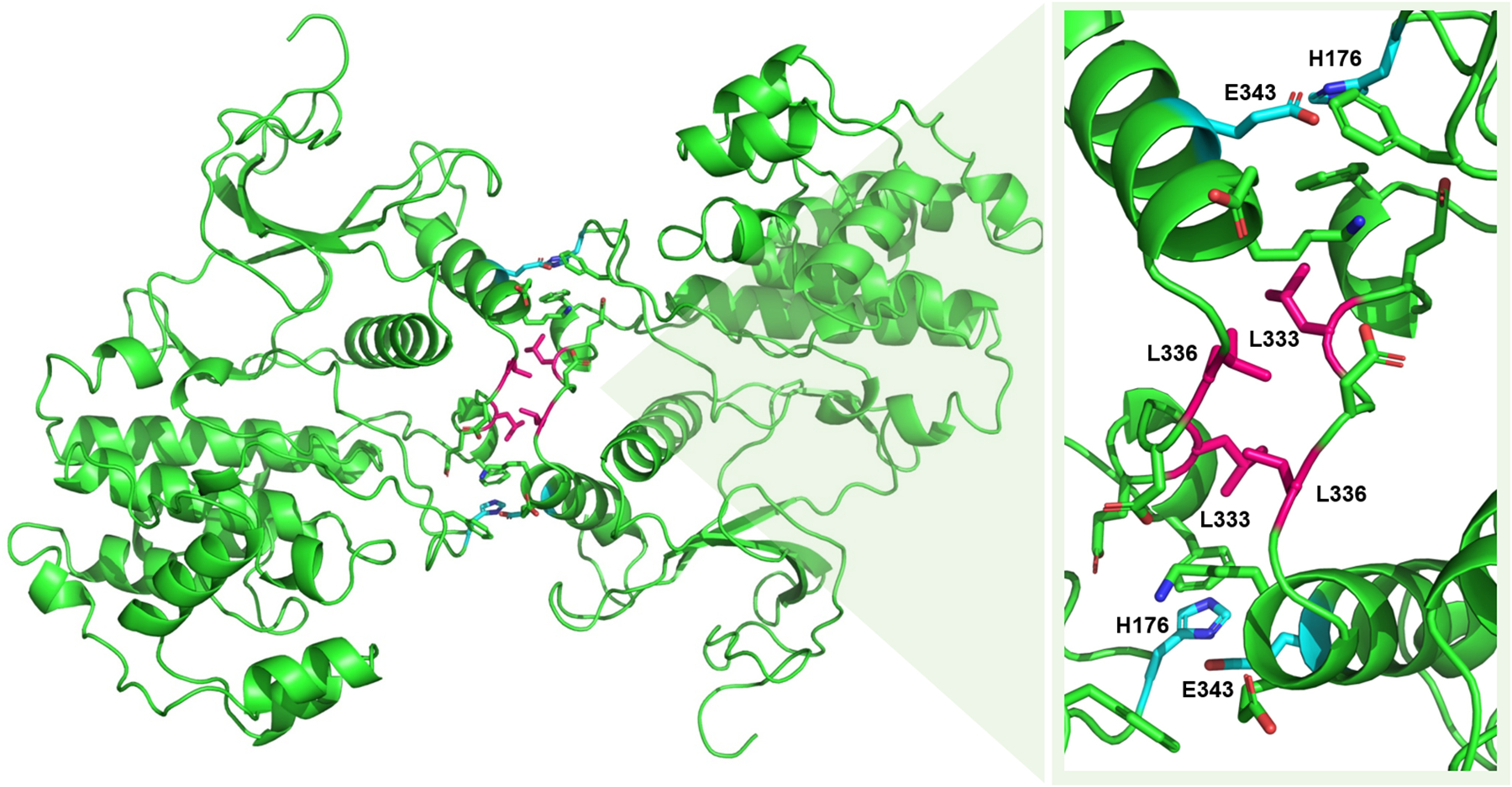
Fig. 5: Active ERK2 homodimer interface.

A crystal of the phosphorylated ERK2 homodimer is shown to the left (PDB ID: 2ERK). A close-up of the dimer interface is shown at the right with key residues labelled. Select residues engaging in leucine zipper-like interactions are shown in pink. The two sets of residues forming a critical salt-bridge are shown in cyan. Figure adapted from Wilsbacher et al. 2006 (79).
