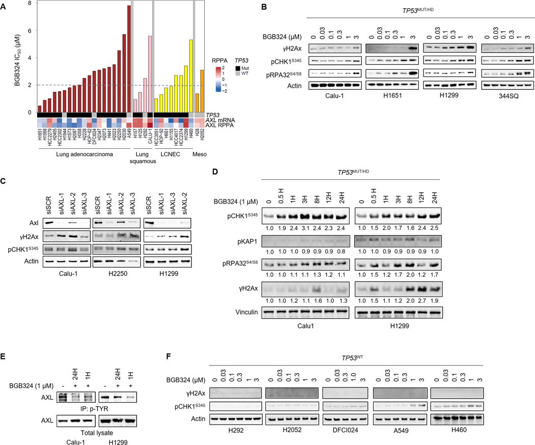
Figure 2. AXL inhibition and knockdown induces DNA damage and activates ATR/CHK1 signaling.
A. Sensitivity of a panel of lung cancer cell lines to BGB324 (ranked by IC50). Dashed line indicates median IC50 (2μM). AXL mRNA levels, AXL protein expression determined by RPPA, and TP53 mutation status are indicated below. B. Expression of markers of DNA damage (γH2Ax) and RS (pCHK1, pRA32) by western blotting, following treatment with indicated concentrations of BGB324 for 24 h, in TP53-deficient lung cancer cell lines. β-Actin was used as loading control. Representative immunoblots of at least 2 independent experiments are shown. C. AXL was silenced in Calu-1, H2250 and H1299 cells using 3 different siRNA sequences. Increase in CHK1 phosphorylation and γH2Ax accumulation 48 h post-AXL depletion was observed by western blotting. D. Calu-1 and H1299 cells were treated with 1μM BGB324 for indicated durations. Expression of the different DDR pathway mediators and Vinculin (loading control) was analyzed by western blotting. Relative band intensities, quantified by Image J and normalized to loading control, are indicated below the blot. E. Inhibition of AXL phosphorylation in Calu-1 and H1299 cells by BGB324. Phosphorylated proteins were immunoprecipitated using Phospho tyrosine antibody and immunoblotted for AXL. F. Immunoblots show limited γH2Ax induction and CHK1 phosphorylation in TP53-wildtype lung cancer cell lines.

