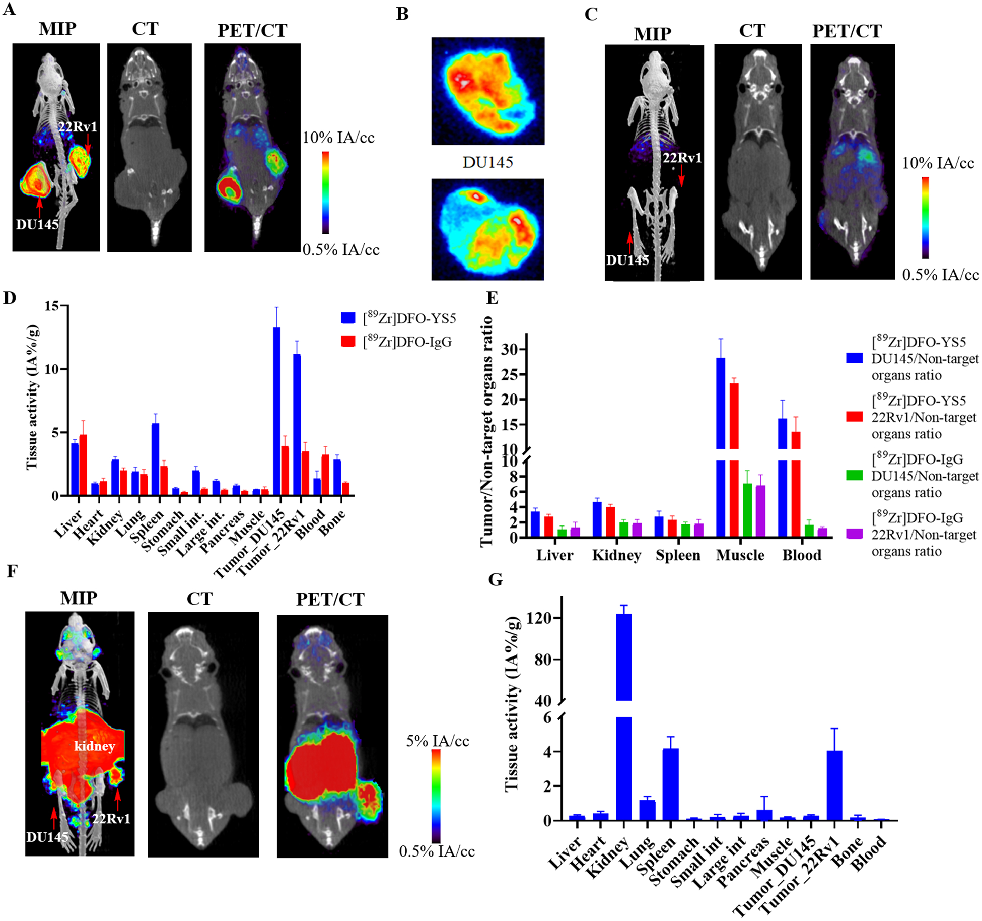
Figure 5:

Comparison of [89Zr]DFO-YS5 and [68Ga]PSMA-11 PET in a dual 22Rv1 and DU145 PCa tumor model demonstrates feasibility for imaging PSMA negative tumors with PET/CT. A) Maximum intensity projection PET/CT, coronal CT and coronal μPET/CT slices obtained 4 days after administration of [89Zr]DFO-YS5 reveal high tumor uptake. B) Autoradiography of 22Rv1 and DU145 tumor sections. C) Maximum intensity projection PET/CT, coronal CT and coronal μPET/CT slices obtained 4 days after administration of [89Zr]DFO-IgG reveal low tumor uptake. D) Biodistribution of [89Zr]DFO-YS5 and [89Zr]DFO-IgG. E) Tissue/Organ ratio of [89Zr]DFO-YS5 and [89Zr]DFO-IgG biodistribution. F) Maximum intensity projection PET/CT, coronal CT and coronal μPET/CT slices obtained 60 minutes after administration of [68Ga]PSMA-11 reveal high tumoral uptake in 22Rv1, but low in DU145. G) Biodistribution data matching imaging data in panel F.
