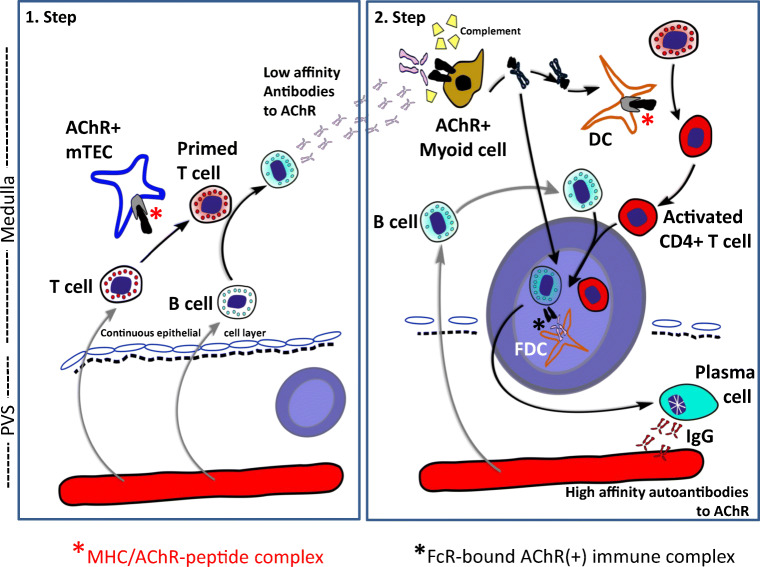
Fig. 3.
Two-step intrathymic pathogenetic model of early-onset myasthenia gravis. Step 1: On re-entry of acetylcholine receptor (AChR)-reactive T-cells from the blood to the thymus, the T-cells (activated by unknown triggers) get ‘primed’ by medullary thymic epithelial cells (mTECs) expressing MHC/AChR-peptide complexes. The primed T-cells activate thymic B-cells to produce low-affinity anti-AChR antibodies. Step 2: These autoantibodies bind to thymic myoid cells (TMCs) expressing native AChRs, activate complement and induce the release of AChR/antibody complexes from TMC for processing by nearby dendritic cells (DCs) that bind to follicular dendritic cells (FDCs). The germinal centre (GC) reaction finally results in plasma cells producing high-affinity anti-AChR autoantibodies. It is unknown whether lymphoid follicles arise primarily in the PVS (as shown on the left and in Fig. 1d) or in the medulla, and why AChR-reactive T-cells occur very commonly in the ‘physiological’ T-cell repertoire of healthy humans

