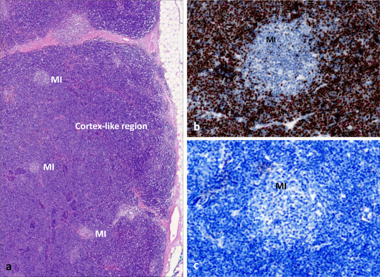
Fig. 4.
Typical abnormalities of a thymoma with extensive thymopoiesis. a Conventional hematoxylin-eosin stain with predominant (dark) cortical areas and tiny (light staining) medullary regions. b TdT stain highlights extensive positively stained cortical areas (C) and small, unstained ‘medullary island’ (MI). c Absence of B-cells throughout the tumour (PAX5 stain). Note absence of Hassall corpuscles (due to absence of AIRE expression, not shown ). Immunoperoxidase

