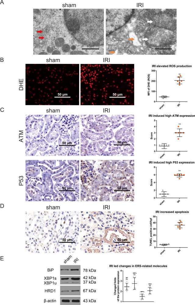
Fig. 1. XBP1 is involved in the crosstalk between mitochondrial damage and ERS in renal IRI.
A Mitochondrial damage and severe ERS occurred simultaneously. TEM analysis revealed mitochondrial swelling, mitochondrial cristae and membrane disappearance, as well as the obvious swelling and vacuolization of ER and detachment of ribosomes from rough ER in IRI group. Normal ER is denoted by red arrows, swelling ER is represented by orange arrows, and swelling mitochondria is indicated by white arrows. Scale bar = 1 μm; ×7000 magnification. B IRI significantly elevated the levels of ROS in IRI group compared to sham group. DHE staining; ×400 magnification; scale bar = 50 μm. **P < 0.01 vs. sham group (n = 6). C The expression of ATM and P53 were raised in IRI group than in sham group. Immunohistochemistry; ×400 magnification; scale bar = 50 μm. **P < 0.01 vs. sham group (n = 6). D The number of apoptotic cells was higher in IRI group than in sham group. TUNEL staining; ×400 magnification; scale bar = 50 μm. **P < 0.01 vs. sham group (n = 6). E The fold changes in the expression of ERS-related molecules in the renal tissues after IRI. The expression levels of BiP, XBP1u, XBP1s, and HRD1 were measured by western blotting. Compared to sham group, the expression levels of ERS-related molecules were all increased in IRI group, especially XBP1s. **P < 0.01 vs. sham group (n = 6).

