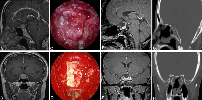
Figure 2.
Reconstruction of the skull base with an autogenous in situ bone flap. (A,B) Preoperative contrast-enhanced MRI of the sellar region indicated that the tumor was to the right, and the structure of the optic nerve was unclear. (C) Formation of the autogenous in situ bone flap intraoperatively. (D) Autogenous in situ bone flap for skull-base reconstruction. (E,F) Three months after the EEEA, MRI of the sellar region indicated that the tumor had totally resected, and that the optic nerve, pituitary stalk and pituitary gland were intact. (G,H) Three months after surgery, CT of the skull base indicated that the bone flap was well restored, and the osseous anatomic structure of the skull base had been restored.

