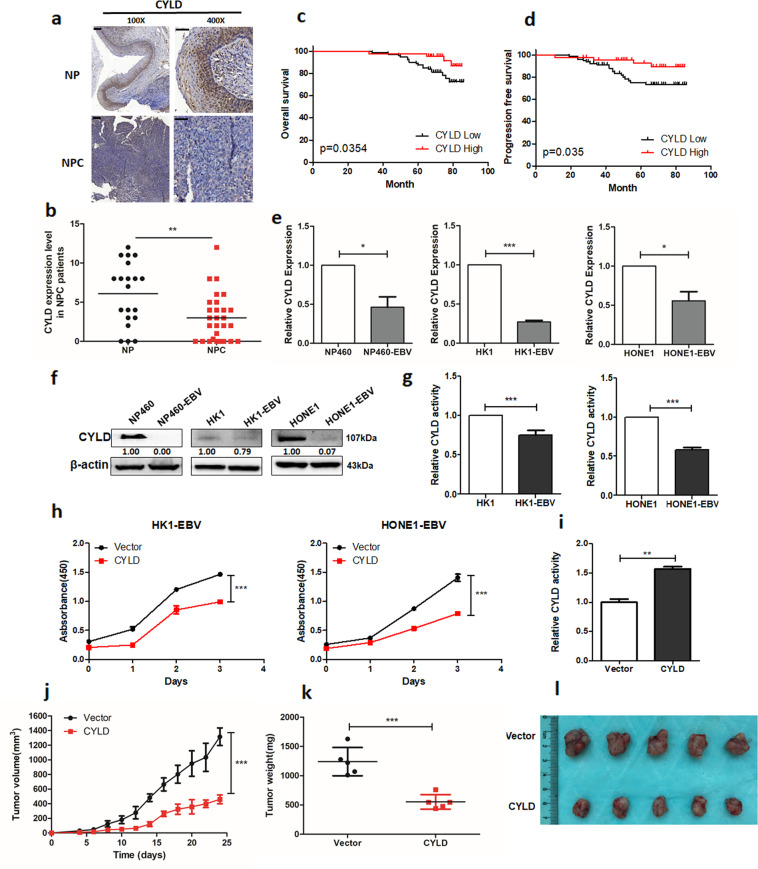
Fig. 1. EBV inhibits CYLD expression and contributes to tumorigenesis in NPC.
a Representative IHC staining of CYLD expression from pathological sections of NPC patients (100×: scale bar, 100 μm; 400×: scale bar, 50 μm). b Histoscores of CYLD expression in NPC patients (n = 29) compared to nasopharyngeal (NP) tissue (n = 20). c Overall survival and d Progression free survival rates of NPC patients with low (n = 71) or high (n = 58) expression levels of CYLD were estimated by the Kaplan–Meier method using log-rank test. Group according to CYLD median expression. e mRNA and f protein expression of CYLD in NP460/NP460-EBV, HK1/HK1-EBV, and HONE1/HONE1-EBV cell lines. g DUB activity measurement of CYLD in HK1/HK1-EBV, HONE1/HONE1-EBV cells. h HK1-EBV and HONE1-EBV cells were infected with a lentivirus and proliferation was monitored using CCK8 assays at the indicated time points. Statistical significance was determined by a two-tailed, unpaired Student’s t test. The indicated cells (5 × 10⁶) were subcutaneously injected into mice. i CYLD activity in tumor extracts. j Tumor growth, k tumor weight, and l tumor images are shown. (Data are represented as mean ± SEM (n = 3). Differences were considered significant at *p < 0.05, **p < 0.001, ***p < 0.001).

