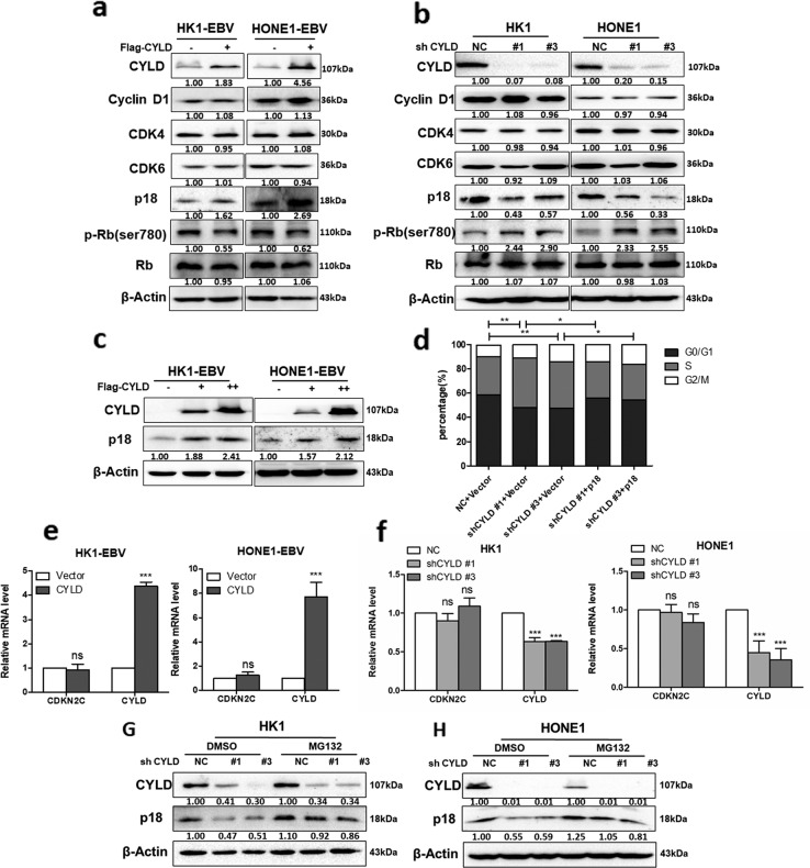
Fig. 3. CYLD regulates the protein level of p18.
a HK1-EBV and HONE1-EBV cells transfected with CYLD were immunoblotted with antibodies against the indicated proteins. b HK1 and HONE1 cells were infected with CYLD lentiviral shRNAs and cell lysates were immunoblotted with antibodies against the indicated proteins. c Increasing amounts of CYLD were transfected into HK1-EBV and HONE1-EBV cells, and total protein was extracted from these cells and subjected to Western blotting using anti-CYLD, anti-p18, or anti-β-actin. d HONE1 cells were infected with the indicated lentiviral shRNAs followed by transfection with the indicated constructs, and then stained with propidium iodide and analyzed by flow cytometry. Total RNA either from cells infected with e CYLD or from cells transfected with the indicated f lentiviral shRNAs was isolated and subjected to qPCR. The error bars represent the S.E.M. of triplicate measurements. g HK1 and h HONE1 cells infected with the indicated lentiviral shRNAs were treated with MG132 (20 μM) for 6 h and the indicated proteins were analyzed by Western blotting. (Data are represented as mean ± SEM (n = 3). Differences were considered significant at *p < 0.05, **p < 0.01, ***p < 0.001).

