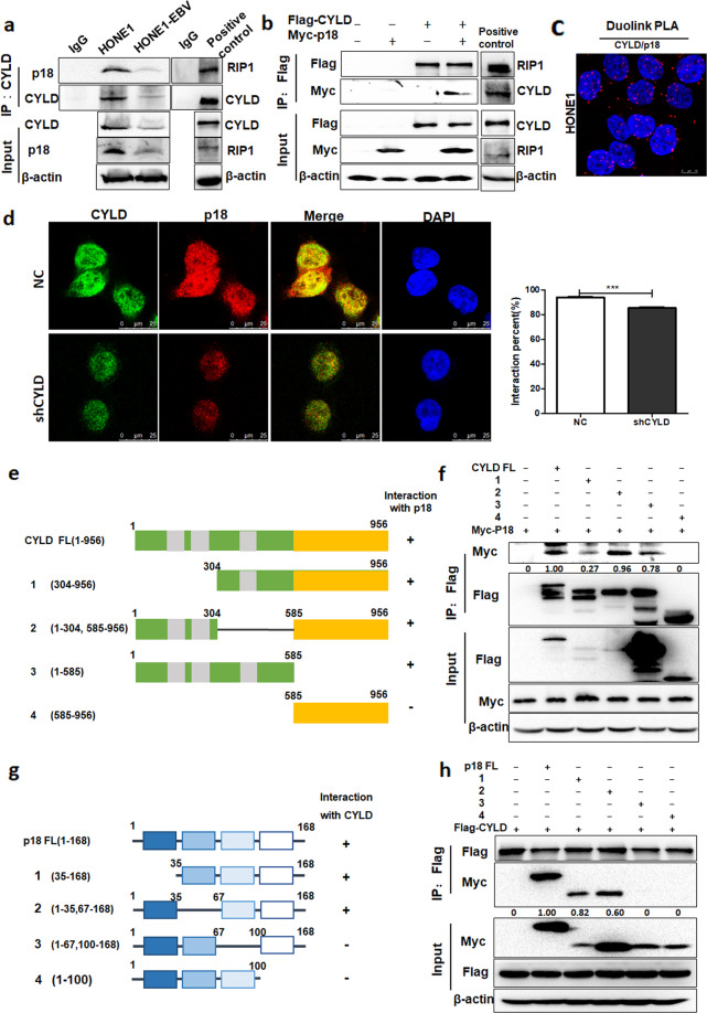
Fig. 5. CYLD interacts with p18.
a Immunoprecipitates were analyzed with CYLD antibodies in HONE1 and HONE1-EBV cells. The arrow points to the destination band. Immunoprecipitates of RIP1 was taken as positive control. b 293T cells were transfected with Flag-CYLD and Myc-p18, and he cell lysates were subjected to immunoprecipitation with Flag antibodies, then the immunoprecipitates were probed with anti-Flag or anti-p18. Immunoprecipitates of Flag-CYLD and Myc-RIP1 was taken as positive control. c Proximity ligation assay indicating the interaction of CYLD and p18 in HONE1 cells (red: PLA positive signal; blue: DAPI, scale bar = 10 μm). d Endogenous CYLD (green) and p18 (red) in HONE1-NC and -shCYLD cells was visualized by immunofluorescence with anti-CYLD and anti-p18. DNA was stained with DAPI, and a merged view of the red and green channels within the same field is shown (merge) (scale bar, 10 μm). The interaction percent was calculated by Image J. e Schematic representation of the Flag-tagged full-length CYLD (FL) and its various deletion mutants, including N-terminal 1-303 deletion (1), Middle domain deletion (2), N-terminal (3) and C-terminal (4) constructs. f 293 T cells transfected with the indicated constructs were disrupted. The cell lysates were subjected to immunoprecipitation with anti-Flag. g Schematic representation of the Myc-tagged full-length p18 (FL) and its various deletion mutants, including N-terminal 1-34 deletion (1), N-terminal 36-66 deletion (2), C-terminal 68-99 deletion (3) and C-terminal 101-168 deletion (4) constructs. h 293 T cells transfected with the indicated constructs were disrupted. The cell lysates were subjected to immunoprecipitation with anti-Flag, and the immunoprecipitates were then probed with anti-Myc and anti-Flag. (Data are represented as mean ± SEM (n = 3). Differences were considered significant at ***p < 0.001).

