Figure 2.
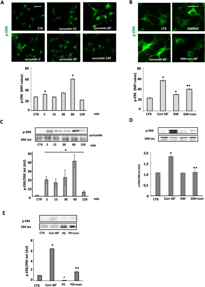
Curcumin modulates phosphorylation of ERK by involving PPAR-γ activation. Immunofluorescence and western blot analysis of the time-course of 1 µM curcumin-induced ERK 1/2 phosphorylation are shown in (A) and (C). Representative photomicrographs of p-ERK immunofluorescence are shown in panel A, at the indicated time points. The upper panel in C shows a representative western blot of p-ERK and total ERK. To confirm the ERK 1/2 activation by curcumin (curc), OPs were pretreated for 30 min with 10 μM PD98059 (PD; ERK1/2 inhibitor) and p-ERK was evaluated by western blot (E). To evaluate the involvement of PPAR-γ in curcumin-induced ERK1/2 activation, OPs were pretreated with 1 μM GW9662 (GW; PPAR-γ antagonist; B and D). p-ERK was evaluated by IF (MFI value is shown in the lower panel in B. Data are mean ± SEM of n. 300 cells for condition) and by western blot (panel D; Data are means ± SEM of three independent experiments). *p < 0.0001 versus CTR; **p < 0.05 vs curc. Scale bar 30 µm. Image Lab 4.0 software (Bio-Rad, http://www.bio-rad.com/en-us/sku/1709690-image-lab-software) and Leica Application Suite Software (260RI, https: //www.leica-microsystems.com/products/microscope-software/details/product/leica-las-x-ls/) were used respectively for Wb and IF image acquisitions.

