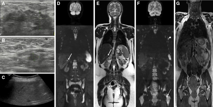
Figure 2.
Pregnant patient with locally advanced breast cancer: breast ultrasound shows (A) large tumorous mass in the left breast with (B) a regional lymphadenopathy in the left axilla. (C) screening ultrasound is equivocal. at whole body diffusion-weighted MRI, (D) the b1000 diffusion-weighted sequence shows a bright lesion in the liver, corresponding to a solid lesion at (E) T2-weighted sequence compatible with liver metastasis (arrows). Additionally, (F) the b1000 diffusion-weighted sequence confirms the lymph node metastasis (arrowheads) as a bright lesion and nodular shape at (G) T1-weighted sequence and shows a bright lesion corresponding to T1 hypo-intensity in the left femur compatible with bone metastasis (dashed arrows).

