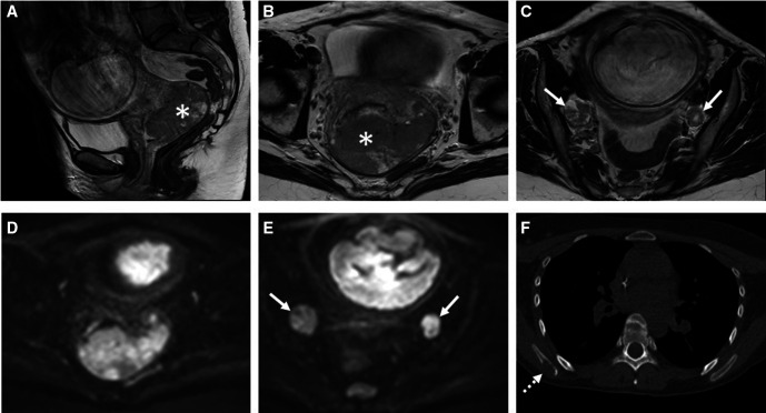
Figure 4.
Pregnant patient with diagnosis of uterine cervical cancer. (A–C) T2-weighted MRI combined with (D, E) diffusion-weighted imaging of the pelvis shows a large tumorous mass (asterisk) in the cervix and prolabating in the proximal vagina without parametrial invasion but with bilateral para-iliac lymphadenopathies (arrows). (F) staging chest CT shows an osteolytic lesion in the right scapula compatible with skeletal metastasis.

