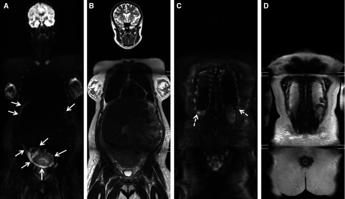
Figure 5.
Pregnant patient diagnosed with a malignant ovarian mass at gynecological ultrasound. (A, C) B1000 whole-body diffusion-weighted MRI shows multifocal and diffuse bright confluent peritoneal and pleural lesions compatible with diffuse metastases. correlative. (B, D) T2-weighted images show thickening of the peritoneal planes, ascites respectively bilateral pleural fluid.

