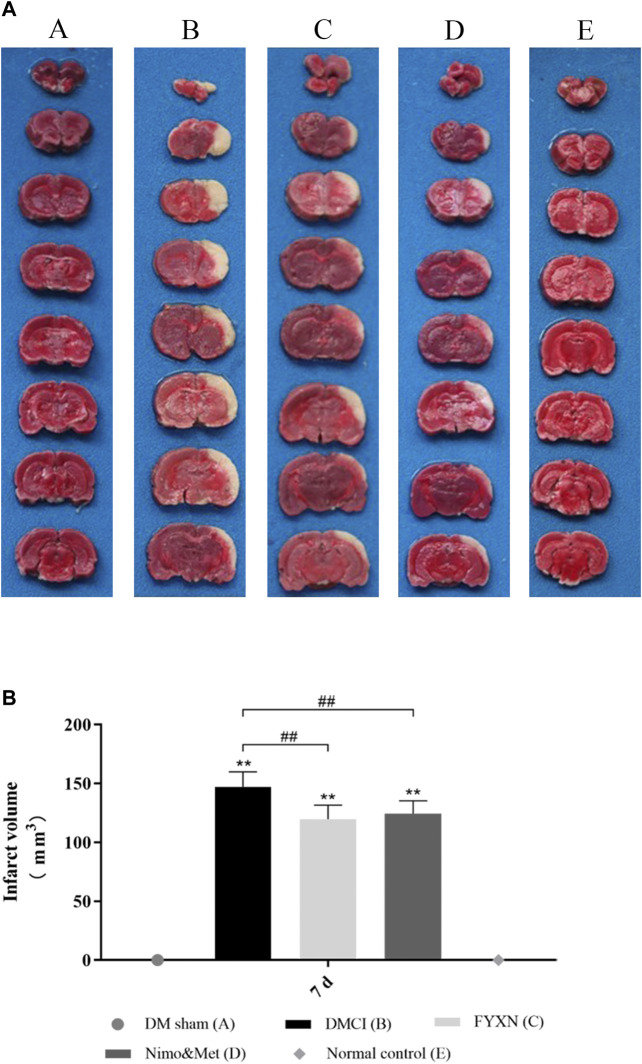
FIGURE 4.
(A) Focal cerebral infarct volume in the experimental groups after 7 days of ischemia followed by 7 days of treatment; 2,3,5-Triphenyltetrazolium chloride staining shows non-infarct (red) and infarct (white) regions. (B) Data of cerebral infarct volume are shown as mean ± SD in each group. *p < 0.05, **p < 0.01 compared with NC group; #p < 0.05, ##p < 0.01 compared with DMCI group.

