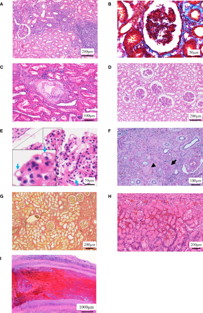
Figure 4.
Histopathological examinations. (A–C) The histopathological images in No. 2 are shown. (A) Localized cell infiltration is observed, and cell infiltration is observed around the epithelial edema. (B) Mild fibrosis is observed (Masson’s trichrome staining). (C) Stenosis of the lumen in the kidney artery, necrosis of the intima and edema of the media, and cell infiltration in the adventitia are observed. (D, E) The histopathological images in No. 3 are shown. (D) Borderline change is observed to the extent that localized cell infiltration is observed. (E) Chronic glomerulosis due to thickening of the vascular loop (blue arrow) is shown with an increase in mesangial substrate. (F, G) The histopathological images in No. 4 are shown. (F) Arterial infarction (arrow) and venous infarction (arrowhead) are observed. (G) Fibrosis was mild but more extensive than No. 2 (Elastica van Gieson staining). (H, I) Representative histopathological images of individuals who died early after operation are shown. (H) Tubular necrosis is observed throughout the whole kidney. Infiltration of mononucleosis is observed near the capsule. (I) Thrombus formation is observed in the renal artery.

