
FIGURE 3:
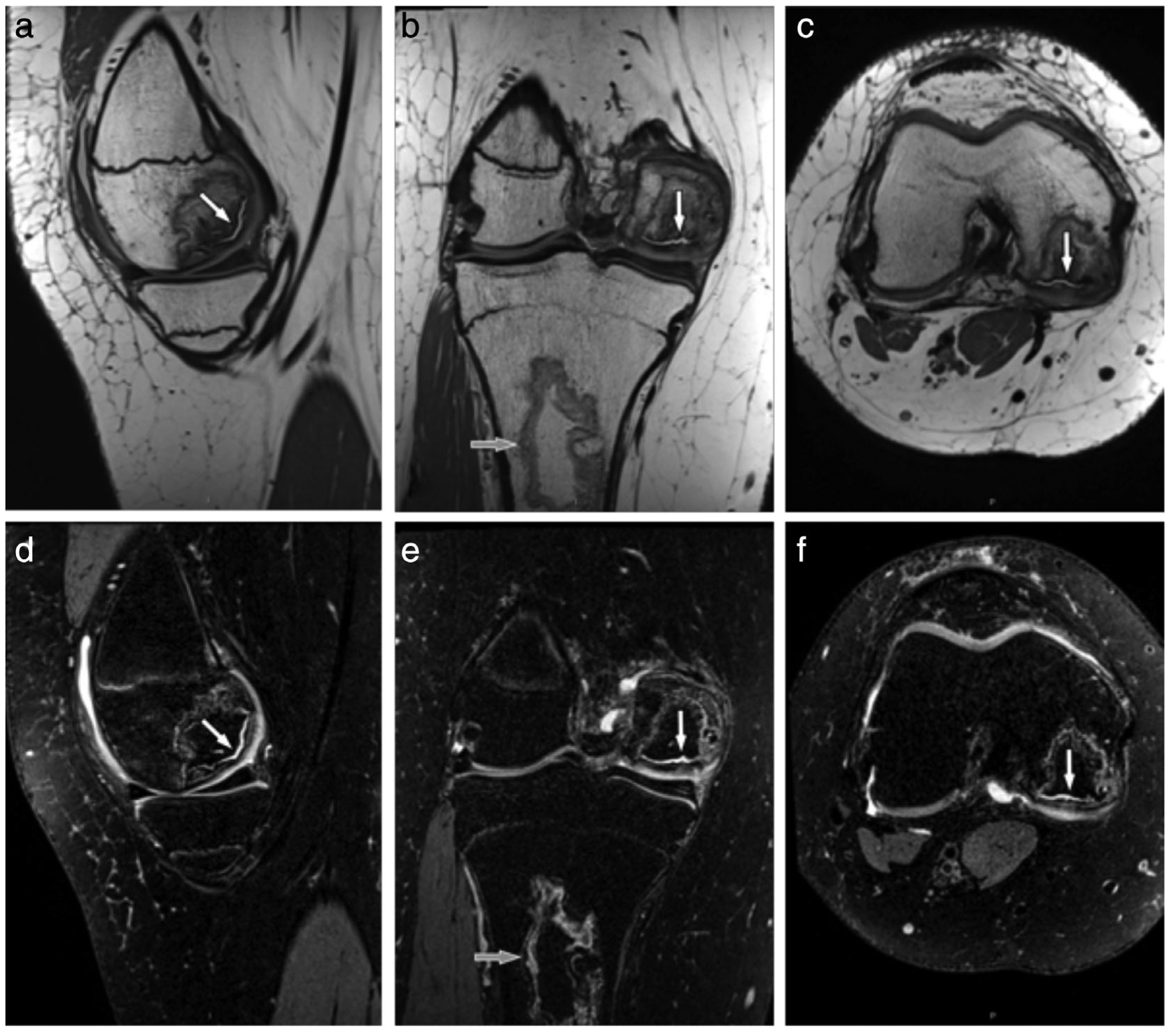
A 16-year-old female teenager with a history of leukemia and long-term glucocorticoid treatment. Isotropic sagittal intermediate-weighted (a) and fat-suppressed T2-weighted (d) CAIPIRINHA (controlled aliasing in parallel imaging results in higher acceleration) SPACE TSE MR images with coronal and axial intermediate-weighted (c,d) and fat-suppressed T2-weighted (e,f) reformation MR images show an osteonecrosis of the medial femoral condyle with separation of a large osteochondral fragment as indicated by intersecting joint fluid (white arrows). In addition, there is a medullary bone infarct (b,e; gray arrows) in the proximal tibial metaphysis. The intermediate-weighted 3D CAIPIRINHA SPACE TSE dataset was acquired with a 0.5 × 0.5 × 0.5 mm3 spatial resolution and a total acquisition time of 4 minute and 41 seconds. The fat-suppressed T2-weighted 3D CAIPIRINHA SPACE TSE dataset was acquired with a 0.6 × 0.6 × 0.6 mm3 spatial resolution and a total acquisition time of 4 minutes and 45 seconds. No interpolations techniques were used. Images courtesy Dr. Jan Fritz, Johns Hopkins University, Baltimore, MD.