
FIGURE 5:
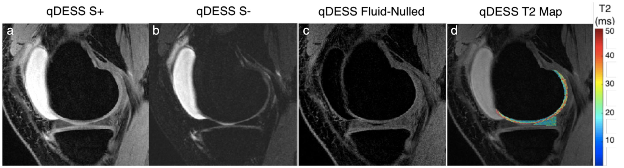
Representative images from a quantitative double-echo steady-state (qDESS) sequence that can generate two novel contrasts. The first echo (S+) generates a T2/T1-weighted contrast (a) while the second echo (S–) has a higher T2 weighting (b). By performing a weighted subtraction of the two echoes, fluid nulling can be performed to enhance visualization of soft tissues (c). Analytical modeling of the two echoes can also be used to accurately characterize the T2 relaxation times of the cartilage and meniscus (d).