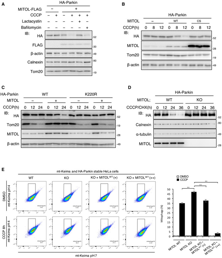
Figure 2. MITOL regulates mitophagy through the degradation of Parkin.

-
AMITOL degrades Parkin through the ubiquitin–proteasome system. HeLa cells stably expressing HA‐Parkin were transfected with the indicated vectors and treated with DMSO or CCCP (10 μM) for 8 h alone or either with bafilomycin A1 (10 μM) or lactacystin (10 μM) for the final 5 h. Lysates of cells were subjected to an IB assay with the indicated antibodies.
-
BOverexpression of MITOL CS mutant fails to degrade Parkin. HeLa cells stably expressing HA‐Parkin were transfected with the indicated vectors and treated with DMSO or CCCP (10 μM) for the indicated time. Lysates of cells were subjected to an IB assay with the indicated antibodies. MITOL CS mutant; lacking ubiquitin ligase activity.
-
CParkin K220R mutant is not regulated by MITOL in mitophagy. HeLa cells were transfected with the indicated vectors and treated with DMSO or CCCP (10 μM) for the indicated time. Lysates of cells were subjected to an IB assay with the indicated antibodies.
-
DEndogenous MITOL attenuates Parkin in both CHX‐ and CCCP‐treated conditions. WT or MITOL KO HeLa cells stably expressing HA‐Parkin were treated with CCCP (10 μM) as indicated times. CHX (30 μM) was added 5 h after CCCP treatment. Lysates of cells were subjected to an IB assay with the indicated antibodies.
-
EMITOL regulates mitophagy. MITOL WT HeLa cells, MITOL KO HeLa cells, and KO HeLa cells transfected with MITOL WT plasmids 0.5 μg (+) and 5 μg (++) stably expressing HA‐Parkin and mt‐Keima were treated with CCCP (10 μM) for 8 h. Then, mKeima was measured at 488 (pH 7) and 561 (pH 4) nm lasers using Flow Cytometer. Percentages of mitophagy were calculated from 30,000 cells in each independent experiment. Data represent the mean ± SD of three independent experiments. For statistical analysis, a one‐way ANOVA with Tukey post‐test was performed, **P < 0.01.
