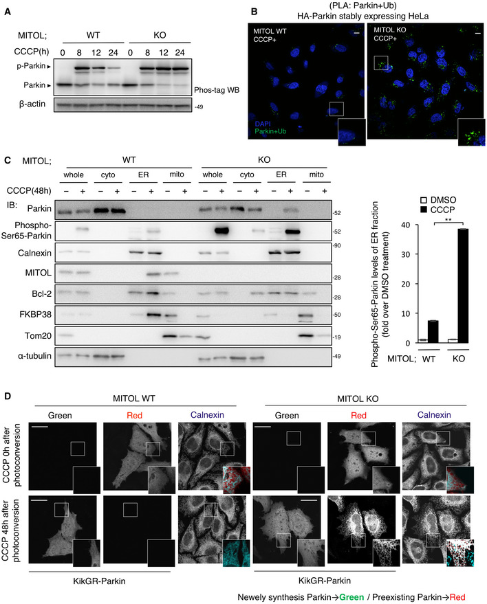
Figure 4. MITOL selectively recognizes phosphorylated form of Parkin.

-
APhosphorylated Parkin accumulates in MITOL KO cells. HeLa cells stably expressing HA‐Parkin were treated with DMSO or CCCP (10 μM) for the indicated time and subjected to IB assay with the indicated antibodies. Other cell lysates were subsequently separated on a Phos‐tag gel, followed by IB with anti‐Parkin antibodies (Phos‐tag). p‐Parkin, phosphorylated Parkin.
-
BAccumulation of activated Parkin in MITOL KO cells. WT or MITOL KO HeLa cells stably expressing HA‐Parkin were treated with CCCP (10 μM) for 30 h and then performed Duolink in situ proximity ligation (PLA) assay demonstrating the interaction between Parkin and Ubiquitin. Scale bar, 5 μm. Higher magnification images of the boxed regions are shown in the small panel.
-
CPhosphorylated Parkin accumulates in the ER fraction in the absence of MITOL. WT or MITOL KO HeLa cells stably expressing HA‐Parkin were treated with DMSO or CCCP (10 μM) for 48 h. Lysates of cells were fractionated into cytosolic, mitochondrial, and ER fractions, and then subjected to an IB assay with the indicated antibodies. Phospho‐Ser65‐Parkin is detected by using specific antibody. Right panels are the quantification of IB about Phospho‐Ser65‐Parkin protein levels of the ER fraction. The data represent the mean ± SD for three independent experiments. For statistical analysis, a one‐way ANOVA with Tukey post‐test was performed, **P < 0.01.
-
DParkin is detected at the ER in the absence of MITOL. HeLa cells were transfected with vectors for KikGR‐Parkin. After ultraviolet light (365 nm) exposure to the whole plate, cells were incubated with or without CCCP (10 μM) for indicated time, and monitored for red and green KikGR fluorescence. Scale bar, 10 μm. Higher magnification images of the boxed regions are shown in the small panel.
