
Figure EV4. LegA5 is not required for the anchoring of SidC to the LCV.
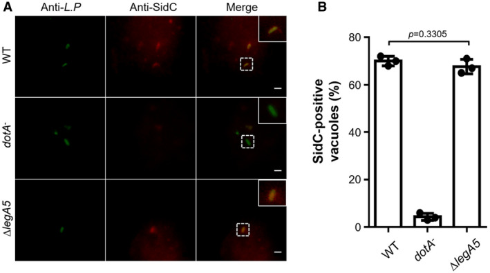
- Representative immunofluorescence images of anti‐Legionella and anti‐SidC staining of bacterial phagosomes. BMDMs were infected with the indicated L. pneumophila stains at an MOI of 2 for 2 h. SidC association on the bacterial phagosomes was detected as described in Fig 5. Insets represent 3× magnification of regions defined by dash lines. Scale bar, 5 μm.
- Percentages of SidC‐positive LCVs. At least 100 phagosomes (n ≥ 100) were scored for each sample. Results are shown as mean ± SD of three independent experiments (biological replicates, n = 3). Unpaired two‐tailed Student’s t‐test, p < 0.05 indicates significant difference.