
Figure 1. Establishing H3K27me3 mintbody to visualise H3K27me3 in living cells.
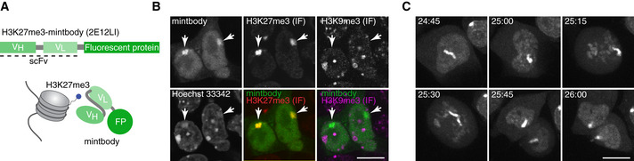
- Schematic representation of the mintbody. H3K27me3‐specific single‐chain variable fragment (scFv) is genetically fused with a fluorescent protein (FP).
- Immunofluorescence (IF) validation of mintbody specificity. Mouse MC12 cells, which stably express H3K27me3‐mintbody (sfGFP), are labelled with antibodies specific for H3K27me3 (Cy5) and H3K9me3 (Cy3). DNA is stained with Hoechst33342. Single confocal sections are shown. Arrows mark Xi. Scale bar = 10 μm.
- Time‐lapse imaging of a dividing MC12 cell stably expressing H3K27me3‐mintbody (sfGFP). Projection images of 7 confocal sections with 2 μm intervals are shown with elapsed time (hh:mm). Scale bar = 10 μm.