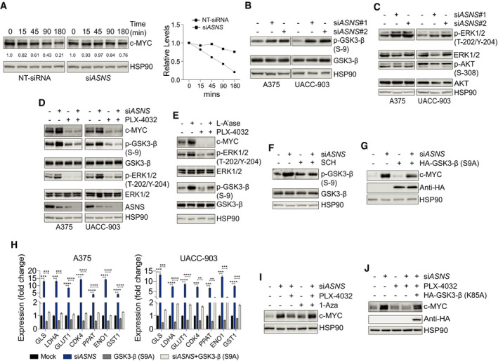
Figure 2. MAPK‐GSK3‐β signaling pathway is required for AAR signaling activation in asparagine‐restricted melanoma cells.

-
AImmunoblotting of c‐MYC in A375 cells treated with NT‐siRNA or siASNS for 24 h followed by cycloheximide treatment for indicated timepoints (left). Plot showing relative protein levels of c‐MYC (y‐axis) as a function of time lapsed (x‐axis) in the left panel (right).
-
B–DImmunoblotting of indicated proteins in melanoma cells 72 h after treatment with NT‐siRNA (−) or siASNS (#1 or #2) (B and C), or siASNS, PLX‐4032, or both (D).
-
EImmunoblotting of indicated proteins in A375 cells cultured in L‐Asn and treated with L‐A’ase, PLX‐4032, or both for 72 h.
-
FA375 cells were treated with siASNS, SCH772984, or both for 72 h followed by immunoblotting of phosphorylated and total GSK3‐β.
-
GA375 cells were treated with siASNS, HA‐GSK3‐β (S9A), or both for 72 h followed by immunoblotting of indicated proteins.
-
HRT–qPCR analysis of indicated transcripts in melanoma cells 36 h after treatment with siASNS, HA‐GSK3‐β (S9A), or both.
-
IA375 cells were treated as indicated for 72 h followed by immunoblotting of c‐MYC.
-
JImmunoblotting of c‐MYC and HA proteins in A375 cells treated as indicated for 72 h.
Data information: Data are shown as the mean ± SD, n = 3 biological replicates. **P ≤ 0.01, ***P ≤ 0.001, ****P ≤ 0.0001. Unpaired Student’s t‐test was used in (H).
Source data are available online for this figure.
