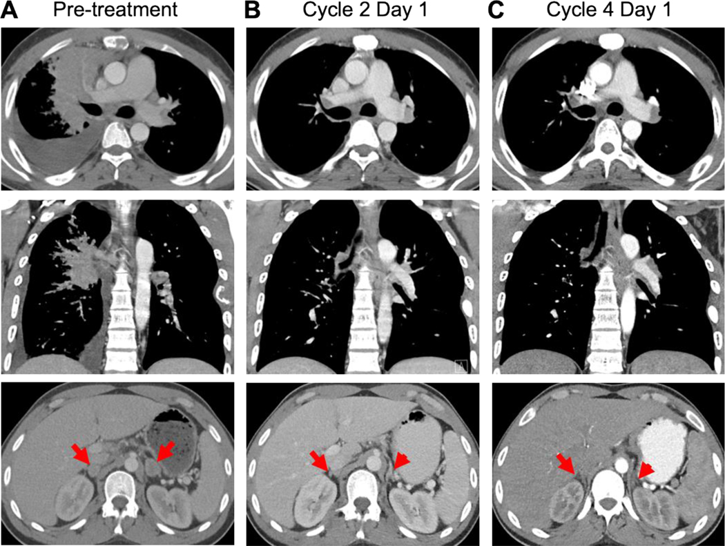
Figure 6. Radiologic response to tarloxotinib.
Baseline imaging obtained prior to dosing with tarloxotinib (150 mg/m2 IV weekly) showed bulky right hilar mass, right pleural effusion, and bilateral adrenal gland lesions (red arrows) (A). A marked tumor response with decreased size of right hilar mass, right pleural effusion, and lesions in the adrenal glands was observed at week 4 (B) and week 12 (C).

