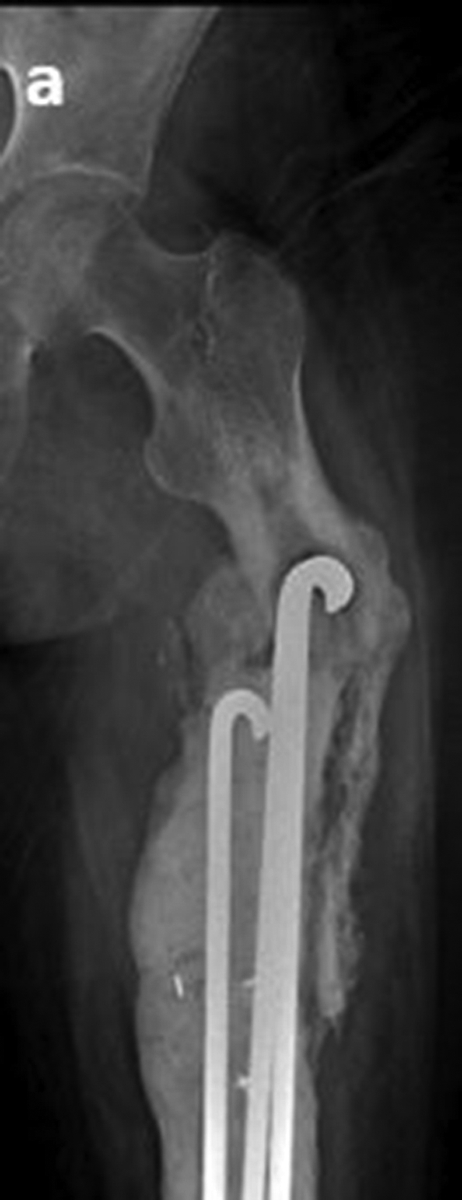
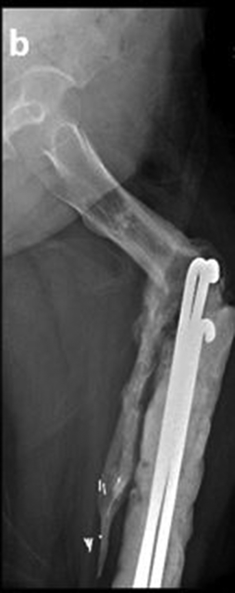
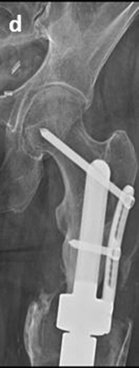
Figure 4.


Secondary reconstruction of a failed, infected distal femoral replacement after osteosarcoma. a. Antero-posterior radiograph shows a pin-cement spacer and malalignment of the short proximal femoral segment. b. Lateral radiograph shows drastic malalignment of the short segment and the spacer. c. Oblique radiograph shows the distal femoral spacer. d. Antero-posterior radiograph shows stable fixation of the stem-side plate 10 years and 7 months after reconstruction. e. Lateral radiograph shows stable fixation 10 years and 7 months after reconstruction.
