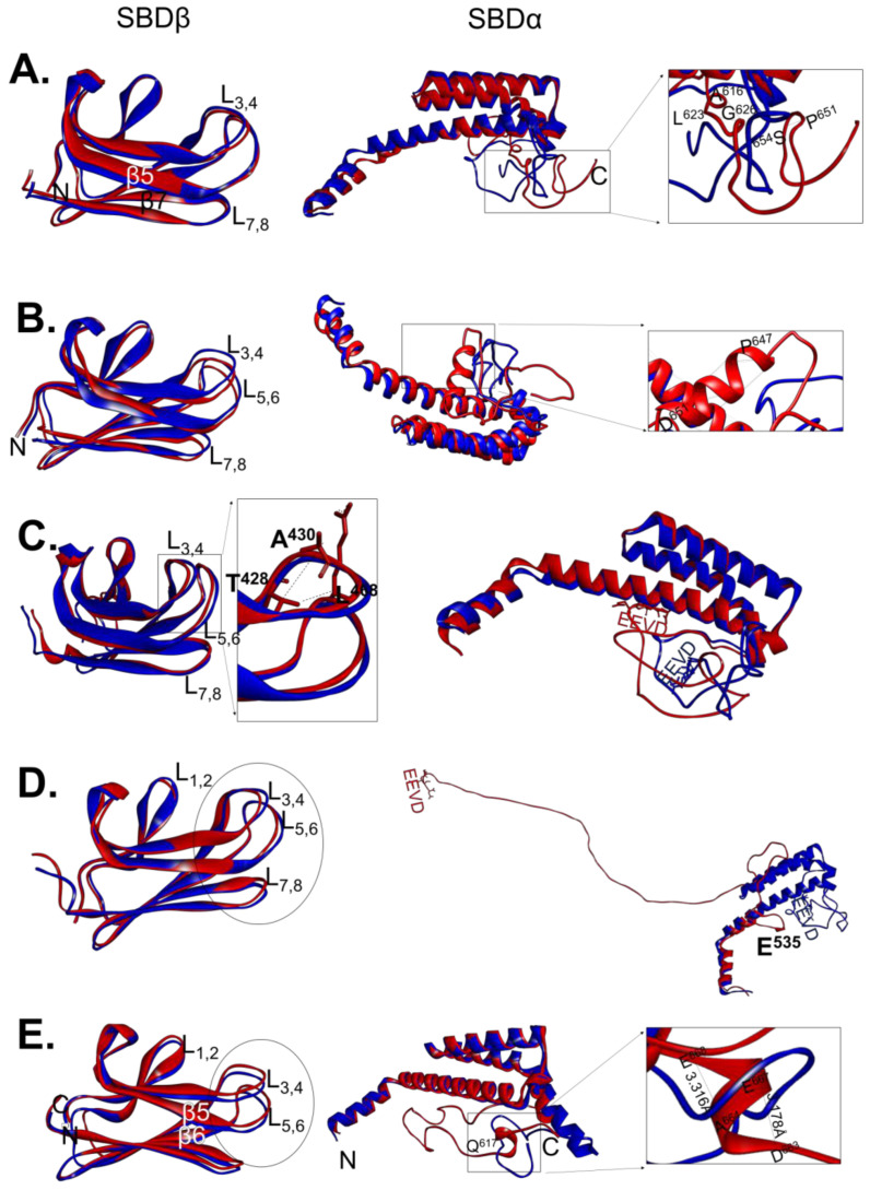
Figure 2.
Superimposed three dimensional models of wild type SBDs of PfHsp70-1 and DnaK versus those of their GGMP variants. Superposition of the SBDβ and SBDα regions of PfHsp70-1 and DnaK against their respective GGMP variants was conducted: (A) PfHsp70-1 versus PfHsp70-1G632, the insert represents zoomed segment highlights H-bonding variations; (B) PfHsp70-1 versus PfHsp70-1G648, the zoomed SBD section with unique H-bonding is shown; (C) PfHsp70-1 and PfHsp70-1G632-664, with the insert showing unique H-bonding variations in SBDβ; (D) PfHsp70-1 versus PfHsp70-1ΔG structures and highlighted are structural variations within the SBD; and (E) DnaK superposed with DnaK-G, insert highlights unique H-bonding pattern. The structures were visualized using Chimera.

