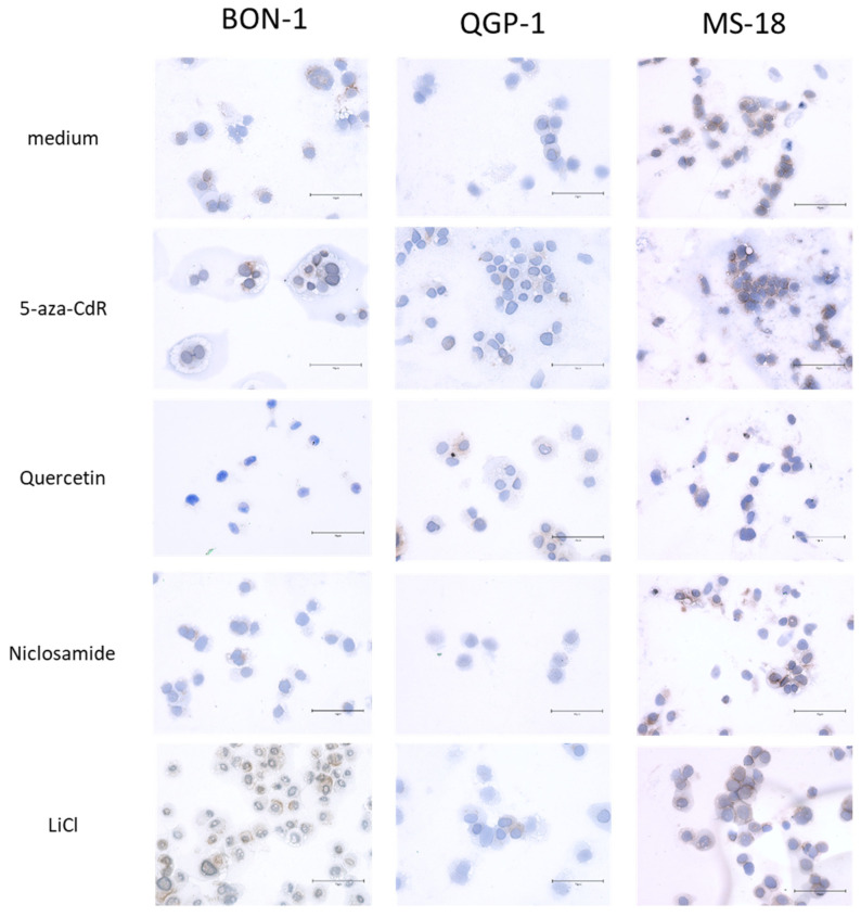
Figure 4.
Immunohistochemistry staining of CXCR4 in BON-1, QGP-1, and MS-18 cells either before (control) or after incubation with Wnt modulators. The bar in the images represents 75 μm. The percentage of CXCR4-positive cells after immunohistochemical staining in BON-1, QGP-1, and MS-18 after Wnt modulator treatment is shown in Supplementary Materials Figure S2.

